Welcome to Class 9 Tissues notes for Chapter 3. The topics in this page are What are tissues, Importance of tissues, Classification of tissues, Plant Tissues, Animal Tissues, Epithelial Tissue, Muscular Tissue, Connective Tissue, and Meristematic Tissues. This is according to CBSE and the NCERT Exploration textbook. If you like the study material, feel free to share the link as much as possible.
A group of cells similar in structure that work together to perform a particular function forms a tissue.
All types of tissues have two basic components:
Cells: having common origin and function.
Inter-cellular substances: Are non-living, fibrous, jelly-like substances (also called matrix).
Importance of tissues
Formation of tissues has brought about division of labour in multicellular organisms.
Tissues become organised to form organs and organs into organ systems.
Workload of individual cells has decreased due to origin of tissues.
As a result of improved organisation and higher efficiency, multicellular organisms have higher survival.
💡 New in Exploration Textbook
Different tissues coordinate with one another to perform complex life processes. For example, in animals, muscle tissue enables movement and nervous tissue carries messages to different parts of the body. In plants, conducting tissues such as xylem transport water and minerals, while phloem transports food.
Plants: Most plants are fixed in one place and do not move. They need support to stay firm and upright. Plant cells have a rigid cell wall that provides rigidity and strength.
Animals: Most animals can move from place to place. Without a rigid cell wall, animal cells can change shape easily. This cellular flexibility eventually helps make their bodies suitable for locomotion.
A meristematic tissue constitutes a group of actively dividing cells present in the growing region of plant, e.g., the tips of roots and stems.
These tissues are responsible for increasing the length and girth of the plant.
Characteristics of meristematic tissues
The cells of the meristematic tissue are similar in structure and have thin cellulose cell walls.
The cells may be spherical, oval, polygonal or rectangular in shape.
The cells of tissue are compactly arranged and do not have intercellular space.
The cells have dense cytoplasm (protoplasm) with prominent nuclei and many organelles.
Vacuoles in these cells are either small or absent.
These cells divide continuously and rapidly.
⚠️ Think About It
Why do you think that the cells of meristematic tissues lack vacuoles?
Show Answer
Vacuoles store water and nutrients, which would take up space needed for the cell's machinery for rapid division. The dense cytoplasm with many organelles allows for continuous cell division without the space taken up by large vacuoles.
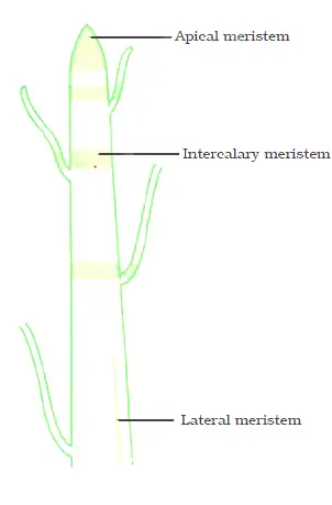
On the basis of their position in the plant body, meristematic tissues are classified into three types:
Apical
Lateral
Intercalary
Apical meristems
These are present at the tips of roots, shoots, branches and leaves.
It brings about the elongation of the root and stem. It results in increase in the height of the plant, which is called primary growth.
Lateral meristems
These are present along the lateral side of the stems and roots. For example: cork cambium.
It causes the organ (stem or root) to increase in diameter and girth. This is called secondary growth.
New in Exploration: The increase in girth occurs due to the presence of actively dividing cells arranged in a ring in the stem. These cells divide and produce new cells inside and outside in a concentric manner, leading to an increase in diameter of stem.
Intercalary meristems
They are located at the base of leaves or internodes (e.g., stems of grasses and other monocots) and below the nodes (e.g., mint).
It produces an increase in length of organ such as leaves and internodes.
New in Exploration: The node is the point on plant stem where branches or leaves arise. The part of stem between the two nodes is called internode.
When the hedge around a garden is cut, after some time more branches appear again, giving the hedge a bushy appearance. Grass also appears after some time being mowed or grazed by animals. This happens because of the presence of intercalary meristem at the nodes of its stem.
Your Image: Meristematic tissue types and locations
📖 Suggested NCERT Figure
NCERT Figure 3.3: Location of apical meristem in a sapling — showing shoot apical meristem at the tip and root apical meristem at the root tip
NCERT Figure 3.5: New branches arising from the node of a stem after cutting
NCERT Figure 3.6: Lawn mowing — showing regrowth of grass
3.1 Activity 3.1: Onion Root Growth and Apical Meristem
Aim: To study the growth of roots in an onion bulb and understand apical meristem
Meristematic tissue acts as a parent tissue from which other tissues develop.
These tissues take part in growth by formation of new cells.
The place of injury in plants is healed up by the formation of new cells by meristems.
They are responsible for all types of growth: increase in length (apical), increase in girth (lateral), and regrowth after cutting (intercalary).
💡 Key Concept: Differentiation
Due to continuous cell division, meristematic tissue adds new cells to the plant body. Some of the newly formed cells remain meristematic while others lose the ability to divide. The cells that lose the ability to divide undergo changes in structure and function, and become permanent tissues. These cells become specialised to perform specific functions, such as support, transport or storage. This process, by which meristematic tissue becomes specialised to perform specific functions, is called differentiation. Meristematic tissue becomes permanent by the process of differentiation.
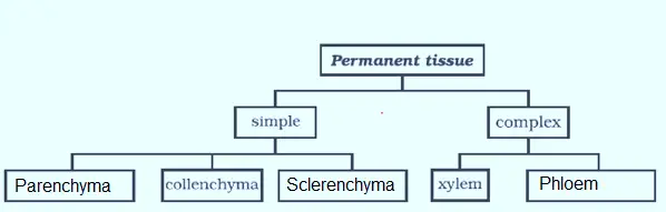
A permanent tissue is a group of cells, which is derived from the meristematic tissues, but these cells have lost the power of division temporarily or permanently.
Note: The development process by which cells which have been derived from meristematic tissue, take up a permanent shape, size and function is called differentiation.
Permanent tissues are of two types:
Simple permanent tissue and
Complex permanent tissue
Your Image: Classification of permanent tissues
📖 Suggested NCERT Figure
NCERT Figure 3.7: Internal structure of a sunflower stem (Transverse Section) — showing epidermis with cuticle, collenchyma, parenchyma, sclerenchyma, xylem, phloem, lateral meristem, ground tissue, and vascular tissue labelled
Simple permanent tissues
These tissues are composed of cells which are structurally and functionally similar. These tissues are of three types:
Parenchyma
Collenchyma and
Sclerenchyma
Parenchyma
Parenchyma forms the bulk of plant body. It consists of thin-walled living cells.
The cells are isodiametric, i.e., equally expanded on all sides.
The cell wall is thin and encloses a dense cytoplasm which contains a small nucleus and a large central vacuole.
The intercellular spaces are abundant (loosely packed).
The parenchyma is present in all the organs of the plants, i.e., roots, stems, leaves, flowers, fruits and seeds.
Functions of Parenchyma
The main function of parenchymatous tissue is storage of food, e.g., starch in the parenchyma of cortex of potato tuber.
Parenchyma forms the framework of all the plant organs and tissues like cortex, pith etc.
Parenchyma serves as packing tissue to fill the spaces between other tissues.
It stores waste materials of plants such as gum, crystals etc.
The intercellular air spaces of parenchyma cells allow gaseous exchange.
If chloroplast is present, the parenchyma tissue is called chlorenchyma and it performs photosynthesis in the green parts of the plants.
In aquatic plants, large air cavities are present in parenchyma to give buoyancy to the plants to help them float. Such a parenchyma type is called aerenchyma. Specialised parenchyma forms air spaces, which help them float.
Collenchyma
Collenchyma is usually found in 3-4 layers beneath epidermis in stem, petioles and leaves of herbaceous dicot plants.
The cells of this tissue are living, elongated and irregularly thickened at the corners.
New in Exploration: Collenchyma consists of living cells with unevenly thickened corners due to pectin (a chemical that gives flexibility like rubber) deposition.
In collenchyma, intercellular spaces are generally absent.
Functions of Collenchyma
It provides the mechanical support, protection, flexibility and elasticity to the plant organs.
It allows easy bending in various parts of the plant (leaf, stem) without breaking.
This tissue provides support and flexibility, allowing parts of the plant like stems and tendrils to bend without breaking.
When cells of collenchyma contain some chloroplasts, they manufacture sugar and starch.
Sclerenchyma
Sclerenchyma cells are dead cells and they are devoid of protoplasm.
They are long and narrow as the walls are thickened due to lignin; such cell walls are called lignified.
The cells of sclerenchyma are closely packed without intercellular spaces.
Cells of sclerenchyma are of two types: fibres and sclereids.
Fibres consist of very long, narrow, thick and lignified cells. Sclereids are irregularly shaped.
This tissue is present in stems, around vascular bundles, in the veins of leaves and in the hard covering of seeds and nuts. Husk of coconut is made of sclerenchymatous tissue.
New in Exploration: Most of these cells are dead, and this tissue is found in stems, leaf veins, and hard coverings of seeds and nuts, such as coconut husk and walnut shell, making them hard and strong (forms the woody structure).
Functions of Sclerenchyma
The sclerenchyma is mainly mechanical and protective in function.
It gives strength, rigidity, flexibility and elasticity to the plant body and, thus, enables it to withstand various strains.
📖 Suggested NCERT Figure
NCERT Figure 3.8: Various types of simple permanent tissues
(a) Parenchyma — thin walls, loosely packed cells
(b) Collenchyma — thick walls at corners
(c) Sclerenchyma — thick lignified walls
🔬 Pause and Ponder
Question: You may have noticed that fibres of coconut husk are hard and brittle, whereas the leaf stalks of coriander are soft and flexible. Can you find out the reason?
Show Answer
Coconut husk fibres are made of sclerenchyma — dead cells with thick, lignified walls that provide hardness and strength. Coriander leaf stalks contain collenchyma — living cells with unevenly thickened corners due to pectin (a flexible chemical), which allows bending without breaking.
Complex Permanent Tissues
The complex tissue consists of more than one type of cell having a common origin. All these cells coordinate to perform a common function.
Complex tissues are of two types: Xylem or wood and Phloem or bast.
Xylem and phloem are both conducting tissues and also known as vascular tissues; together both of them constitute vascular bundles.
Xylem
Xylem is a vascular and mechanical tissue.
Xylem is composed of cells of four different types:
Tracheids
Vessels or tracheae
Xylem parenchyma
Xylem sclerenchyma (or fibres)
Except xylem parenchyma, all other elements are dead and bounded by thick lignified walls.
Tracheids and vessels are tubular structures and thick-walled.
New in Exploration: Xylem parenchyma are the only living component of xylem while tracheids, vessels, and xylem fibres are primarily sclerenchymatous (dead cells).
Functions of Xylem
The main function of xylem is to carry water and mineral salts upward from the root to different parts of shoots, hence also called water-conducting tissue.
Since walls of tracheids, vessels and sclerenchyma of xylem are lignified, they give mechanical strength to the plant body.
The parenchyma stores food and helps in the sideways conduction of water.
It also provides strength to the plant.
Phloem
Phloem (bast) is a living conducting tissue. It also contains tubes just like xylem but does not perform mechanical function.
Phloem is composed of following four elements or cells:
Sieve tubes
Companion cells
Phloem parenchyma
Phloem fibres
Sieve tubes are slender, tube-like structures with perforated walls. Some cells are long and tubular, joined end to end by perforated walls. These cells form sieve tubes.
Companion cells are living parenchymatous cells lying on the sides of the sieve tubes.
Sieve tube and companion cells have close cytoplasmic connection with each other through fine pits.
New in Exploration: The cellular functions of the sieve tube cells are regulated by companion cells. Companion cells are specialised parenchyma cells. Main function of companion cells is to monitor loading and unloading of sugars in sieve tubes.
Phloem fibres are thick-walled fibres with simple pits. They are primarily sclerenchymatous and provide strength.
Phloem parenchyma are thin-walled, living cells of parenchyma of phloem. They store food materials, and resin, tannins and latex.
New in Exploration: Unlike the xylem, phloem is mostly made up of living cells.
Function of Phloem
Phloem transports (conducts) photosynthetically prepared food materials from the leaves to the storage organs and later from storage organs to the growing regions of the plant body.
Sieve tubes transport food from leaves to other parts of the plant.
📖 Suggested NCERT Figure
NCERT Figure 3.9: Vascular tissue
(a) Xylem showing tracheid, vessel, xylem parenchyma, and xylem fibre
Protective tissues are a part of plant tissue system. Protective tissues include:
Epidermis and
Cork
Epidermis
It is the outermost protective layer of plant organs.
The epidermis is usually made of a single layer of cells.
New in Exploration: The epidermis forms the outermost layer of the plant body. It consists of a tightly packed, single layer of flat and rectangular cells. It protects all parts of the plants.
Cells of epidermis are elongated and flattened, without intercellular space. They are living cells but their inner contents are similar to parenchyma cells.
These cells are covered with a waxy layer of cutin called cuticle.
In some plants living in very dry habitats, the epidermis may be thicker (covered by a thick layer of cuticle) since protection against water loss is critical to reduce the water loss in the process of transpiration by stomata.
The cuticle also provides protection against mechanical injury and invasion by parasites.
In many plants, hair-like projections arise from epidermal cells. In roots, these projections are called root hair, which increase the surface area for absorption of water and minerals from the soil.
In leaves, epidermis bears small pores called stomata.
In leaves, the epidermis contains pores called stomata, which apart from gaseous exchange help in transpiration, i.e., evaporation of water vapours through stomata. Thus, transpiration helps in water transportation by creating a transpiration pull in xylem. Transpiration also helps in elimination of wastes from the plant body.
Functions of Epidermis
The function of epidermis is the protection of plant from injury and infection.
Cuticle of epidermis also helps to reduce water loss by evaporation to prevent desiccation.
Stomata present in the epidermis allow gaseous exchange to occur during photosynthesis and respiration.
It also facilitates transpiration.
Epidermis helps in the absorption, secretion and movement of substances.
Cork (or phellem)
Cork cells are dead cells without having intercellular spaces.
They appear at the periphery of roots and stems when they grow older and increase in girth.
They also have a chemical called suberin in their walls that makes them impervious to gases and water.
New in Exploration: Cork cells are dead, compactly arranged, and contain suberin which makes them impermeable to water and gases.
Functions of Cork
The function of cork in plant body is to provide protection. It protects plants from external injury and infection.
It also prevents desiccation.
Since cork does not catch fire easily, it is used for insulation, shock-absorber, linoleum.
It is also used for making sports goods, such as shuttlecock, table tennis paddles, cricket balls, etc.
📖 Ready to Go Beyond (New in Exploration)
In young plants, the outer protective layer is a single-layered epidermis. As plants grow older, some cells below the epidermis of the stem develop the ability to divide, act as lateral meristematic cells and form the cork cambium. The division of cork cambium cells gives rise to cork cells. Cork cells are dead, compactly arranged, and contain a substance which makes them impermeable to water and gases. This forms the bark of the tree.
🔬 Pause and Ponder (New in Exploration)
Question 1: Why do you think that a thick cuticle on the outer wall of epidermis is advantageous for a plant living in the desert but disadvantageous for a plant living underwater?
Show Answer
Desert plants: Thick cuticle is advantageous because it prevents excessive water loss through transpiration in hot, dry conditions.
Underwater plants: Thick cuticle is disadvantageous because it would prevent gaseous exchange (CO₂ and O₂) with the surrounding water, which is essential for photosynthesis and respiration.
Question 2: Once water is absorbed by plant roots, it has to travel against gravity through xylem. How do the 'dead' cells of the xylem work together with the living cells of leaves at the top to keep the water moving?
Show Answer
The dead xylem cells (tracheids and vessels) form continuous hollow tubes from roots to leaves. Living leaf cells lose water through stomata during transpiration, creating a "pull" or suction force. This transpiration pull draws water upward through the xylem vessels. The cohesion of water molecules and adhesion to xylem walls help maintain the continuous water column against gravity.
Question 3: What do you think will happen if there were no stomata in the epidermis of the stem or leaves?
Show Answer
Without stomata:
• No gaseous exchange — photosynthesis would stop (no CO₂ entry)
• No transpiration — water transport would be severely affected
• No oxygen release — plant respiration would be disrupted
• The plant would eventually die due to inability to perform essential life processes
💡 Plant Tissue Systems (New in Exploration)
In a plant body, different tissues do not work alone. They are organised together into larger groups called tissue systems. Plant tissues are organised into three tissue systems:
Dermal tissue system: This forms the outer covering of the plant. It protects the inner parts and reduces water loss.
Ground tissue system: This forms the main body of a plant between the dermal and conducting tissues. It includes parenchyma, collenchyma and sclerenchyma.
Vascular tissue system: This consists of conducting tissues — xylem and phloem.
NCERT Figure 3.10: Tissue systems in plants — showing cross-sections of different plant parts with epidermal tissue, ground tissue, xylem, and phloem labelled
New in Exploration: Like plants, animal cells also group together and specialise in performing different functions. These groups of similar cells form animal tissues.
Think about this: Which tissue helps you move? Which tissue enables you to sense heat or cold? Which tissue allows oxygen to enter the blood? Which tissue holds the body together so that the skin does not fall off?
Many such questions can be asked but the answers lie in the diversity of animal tissues, which are specially adapted to perform different functions. It is interesting to understand how the structure of an animal tissue suits its specific function.
On the basis of the structure of cells and their function, animal tissues are classified into four major types:
The covering or protective tissues in the animal body are epithelial tissues.
New in Exploration: Epithelial tissue forms the outer covering of the body (skin) and also lines the internal organs, such as the mouth, lungs, blood vessels and intestine.
The cells of this tissue are tightly packed and it forms a continuous sheet. Indeed cells of epithelium contain very little or no intercellular matrix.
New in Exploration: It is composed of closely packed cells with very little space between them. This structure prevents the entry of the germs, reduces water loss, and also helps in the absorption, secretion and movement of substances.
The skin and lining of buccal cavity, blood vessels, alveoli of lungs and kidney tubules are made of epithelial tissue.
Epithelial cells lie on a delicate non-cellular basement membrane which contains a special form of matrix protein, called collagen.
Functions of Epithelial Tissue
Epithelial cells protect the underlying cells from mechanical and chemical injuries and bacterial or viral infection.
It covers most organs and cavities within the body. It also forms a barrier to keep different body systems separate.
Epithelial tissues help in absorption of water and nutrients.
Epithelial tissues help in elimination of waste products.
Some epithelial tissues secrete secretions, such as sweat, saliva etc.
Note: Epithelial tissue may be simple, i.e., composed of a single layer of cells, or stratified, i.e., made up of several layers of cells.
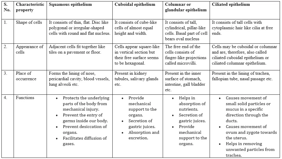
Types of epithelial tissue
Depending upon the shape and function of the cells, the epithelial tissues are classified as follows:
Squamous epithelium
Cuboidal epithelium
Columnar epithelium
Glandular epithelium
Ciliated epithelium
Differences between different types of epithelial tissues
Your Image: Different types of epithelial tissues with structure, location, and function
📖 Suggested NCERT Figure
NCERT Figure 3.11: Types of epithelial tissues in different parts of the body
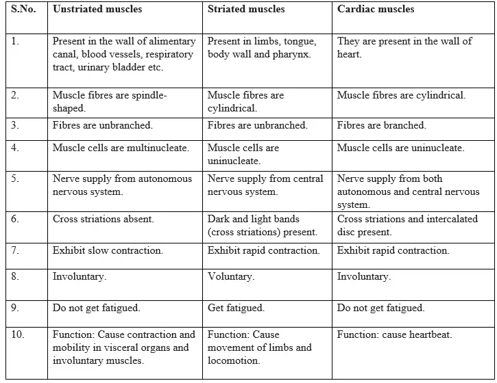
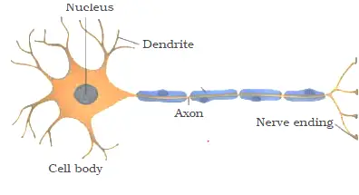
Muscular tissue constitutes all the muscles of the body of an animal.
Muscle cells are elongated and large sized, so they are called muscle fibres.
Muscle cells are typically arranged in parallel arrangement allowing them to work together effectively.
This tissue is responsible for movement in our body. Muscles contain special proteins called contractile proteins (actin and myosin), which contract and relax to cause movement.
On the basis of their location, structure and function, there are following three types of muscle fibres:
Striated muscles (striped, skeletal or voluntary muscles)
Smooth muscles (unstriated, visceral or involuntary muscles)
Cardiac muscles
Your Image: Three types of muscular tissues — Striated, Smooth, and Cardiac
⚠️ Remember (New in Exploration)
Why can cardiac muscle contract continuously without fatigue?
Cardiac muscle cells have a high number of mitochondria (powerhouses) and receive an abundant blood supply, which provides continuous oxygen and nutrients for energy production. This allows the heart to beat continuously throughout life without getting tired.
The connective tissue is specialised to connect and anchor various body organs. As such, it connects one bone with another and a bone with a muscle.
New in Exploration: You have read that blood connects different parts of the body by transporting nutrients, gases, hormones, etc. In the same way, bones connect and support the body from head to toe. A tissue that connects and supports other tissues is called a connective tissue.
Three components are present in all the connective tissues. These are intercellular medium, connective tissue cells and fibres.
The cells of connective tissue are loosely spaced and embedded in an intercellular matrix. The matrix may be jelly-like, fluid, dense or rigid.
The nature of matrix decides the function of connective tissue.
New in Exploration: Both blood and bones are connective tissues. Though both are connective tissues, they differ in composition and consistency. Blood is fluid, while bone is hard. This difference is due to the matrix, which is watery, soft and jelly-like in blood but hard, solid, and rigid in bones.
General functions
Connective tissue binds other tissues together in the organs.
Connective tissue also provides the structural framework and mechanical support to different tissues.
It is also concerned with body defence, fat storage, repair etc.
The main functions of connective tissue are binding, supporting and packing together different organs of the body.
Types of connective tissue
In animals, there are following five types of connective tissues:
Areolar (loose) connective tissue
Dense connective tissue
Adipose connective tissue
Skeletal tissue
Fluid connective tissue
Areolar (loose) connective tissue
It is a loose and cellular connective tissue. Its matrix consists of two kinds of fibres: white collagen fibres and yellow elastic fibres.
Areolar connective tissue is found between the skin and muscles, around blood vessels and nerves and in the bone marrow.
It fills the spaces between different tissues and organs, hence called packing tissue.
Functions of Areolar tissue
It acts as supporting and packing tissue between organs lying in the body cavity.
It provides rapid diffusion of oxygen and nutrients from blood vessels.
It helps in repair of tissues after an injury.
It helps in fighting foreign antigens and toxins.
Dense connective tissue
It is a fibrous connective tissue. It is characterised by ordered and densely packed collection of fibres and cells.
It is the chief component of ligaments and tendons.
Ligaments: These are elastic structures made up of yellow elastic fibrous tissues which connect bone to another bone. It has considerable strength. Ligaments contain very little matrix. Ligaments strengthen the joint and they permit normal movement but prevent over-flexing or over-extension. Sprain is caused by excessive pulling (stretching) of ligaments.
Tendons: Tendons are cord-like, strong inelastic structures that join skeletal muscles to bones. They are composed of white collagen fibrous tissue.
It has great strength but its flexibility is limited.
Adipose tissue
It consists of large number of oval and rounded adipose cells (adipocytes) filled with fat globules.
The adipose tissue is abundant below the skin, between the internal organs (e.g., around the kidney) in yellow bone marrow.
Functions of Adipose tissue
It serves as a fat reservoir.
Adipose tissue acts as food reservoir by storing fat.
It acts as an insulator and regulates body temperature.
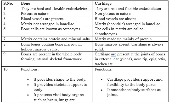
Skeletal tissue
Skeletal connective tissue forms the endoskeleton of the body of vertebrates. It includes cartilage and bone.
Your Image: Skeletal tissues — Cartilage and Bone structure
Fluid connective tissue
Fluid connective tissue links the different parts of the body and maintains continuity in the body. It includes blood and lymph.
Blood
Blood is fluid connective tissue. In this tissue cells move in a fluid or liquid matrix or medium called blood plasma.
The blood plasma does not contain protein fibres but contains cells called blood corpuscles or blood cells. These blood corpuscles and cells are:
Red blood corpuscles (RBC) or erythrocytes
White blood corpuscles (WBC) or leucocytes
Platelets
RBCs and WBCs are living, while plasma and platelets are non-living.
📖 Suggested NCERT Figure
NCERT Figure 3.12: Types of connective tissues in different parts of the body
(a) Blood — fluid matrix with RBCs, WBCs, platelets
(b) Bone — hard, rigid matrix
(c) Cartilage — flexible matrix
(d) Areolar — loose tissue
(e) Adipose — fat storage
3.3 Activity 3.3: Connective Tissues Exploration
Aim: To observe different types of connective tissues and understand their structure
In our body, movement occurs by the coordination of muscles and bones (musculoskeletal system), under the control of the nervous system. The skeletal system provides structure and protection, while muscles produce movement by pulling on bones.
🔬 Bridging Science and Society (New in Exploration)
Crown Gall Disease — From Problem to Solution
In nature, plant pathologists observed a disease in plants called crown gall disease, where tumour-like swellings develop on stems due to rapid and uncontrolled cell division. The disease is caused by the bacterium Agrobacterium tumefaciens.
Instead of only trying to cure this disease, scientists studied how the bacterium transfers its genetic material into plant cells. This knowledge was later used in plant tissue culture and genetic engineering. Today, Agrobacterium is used as a tool to introduce useful genes into plants for the production of valuable phytochemicals, improved crops, and disease-resistant varieties.
Summary
Here is the Tissues class 9 notes Summary:
Tissue is a group of cells similar in structure and function.
Plant tissues are meristematic or permanent.
Meristematic tissue is the dividing tissue present in the growing regions of the plant.
Meristematic tissue becomes permanent tissue when it stops dividing. Permanent tissues are classified as Simple and complex tissues.
Simple tissues include parenchyma, collenchyma, and sclerenchyma. Xylem and phloem are complex tissues.