Coordination is a process through which two or more organs interact & compliment the function of one another.
The neural & endocrine system together co – ordinate & integrate all the activities of the organs.
Neural System in Animals
Sponges do not have neurons.
Hydra (Coelenterates) have similar neurons that form a network.
In earthworm, the nervous system consists of single ventral nerve cord & paired ganglia & segmental nerves forms the segmental ganglia.
Insects have better organized nervous system with brain, ventral nerve cord, ganglia & nerves.
Vertebrates have a well-developed brain.
HUMAN NERVOUS SYSTEM
(Derived from ectoderm)
The human nervous system can be divided as follows:-
Central Nervous System (Brain and Spinal cord)
Peripheral Nervous System (all nerves)
The nerve fibres of Peripheral Nervous System (PNS) are of two types:-
Afferent Fibres:-
They transmit impulses from tissues/ organs to Central Nervous System (CNS).
Efferent Fibres:-
They transmit impulses from CNS to the concerned peripheral tissues/ organs.
SOMATIC NERVOUS SYSTEM:-
It sends impulses from CNS to skeletal muscles.
AUTONOMOUS NERVOUS SYSTEM:-
It sends impulses from CNS to involuntary organs & other smooth muscles of the body.
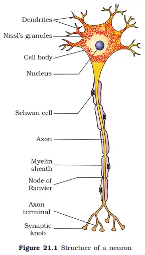
NEURON
Neurons are specialized cells which can detect, receive, transmit different types of stimuli. They are the structural & functional unit of the nervous system.
Each neuron has a cell body (cyton), dendrons (dendrites) and axon. Cell body contain cytoplasm with the nucleus and the nissl's granules (granular bodies).
A number of processes arise from the cell body; the longest among them is called axon, while the others are called Dendrons & their branches are called dendrites.
The axon is a single long fibre & is branched at its distal end & each branch terminates as a bulb like structure called synaptic knob which contains synaptic vesicles with neurotransmitter.
Types of Neuron:
There are two types of neuron on the basis of Myelin sheath:
Myelinated:- The myelinated nerve fibres are enveloped with Schwann cells which form myelin sheath around the axon. The gaps between two adjacent myelin sheaths are called nodes of Ranvier. They are found in cranial & spinal nerves.
Non – myelinated/ Unmyelinated:- In these the Schwann cells do not form myelin sheath. They are found in autonomous & somatic neural system.
Types of Neuron on the basis of structure:-
Based on the no. of axon & dendrites the neurons are divided into three types:-
Multipolar Neuron:-
These have several dendrites & one axon. They are found in the cerebral cortex.
Bipolar:-
These have 1 dendrite & 1 axon & present in the retina of the eye.
Unipolar:-
These have a cell body with 1 axon only & found usually in the embryonic stage.
Main Properties of Neural Tissues:-
Excitability
Conductibility
Transmission of impulses across the synapses
There are two types of synapses namely:-
Electrical synapses
Chemical synapses
Electrical synapse
Chemical synapse
The membranes of the pre synaptic & post synaptic neurons are in close proximity and there is no synaptic cleft.
The membranes of pre- synaptic & post synaptic are separated by a fluid filled space, the synaptic cleft.
Electrical current can flow directly from one neuron to the other.
Transmission involves chemicals called neurotransmitters.
Impulse conduction is faster.
Impulse conduction is relatively slow.
Electrical synapses are rare in our system.
Chemical synapses are the most common types of synapses.
MECHANISM OF SYNAPTIC TRANSMISSION(chemical synapse)
In the chemical synapses, the axon terminal called synaptic knob, contains a number of synaptic vesicles, which contain the neurotransmitter.
When an impulse arrives at the axon terminal, it stimulates the movement of the synaptic vesicles towards the membrane, where they fuse with the membrane & release the neurotransmitter into the synaptic cleft.
The neurotransmitter molecules diffuse across the synaptic cleft & bind to specific receptors present on the postsynaptic membrane.
This binding stimulates the opening of ion –channels & a new action potential is generated in the post – synaptic neuron.
GENERATION & CONDUCTION OF NERVE IMPULSE
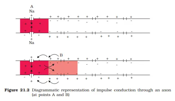
Nerve impulse is a wave of bioelectric/ electrochemical disturbance that passes along a neuron during conduction of an excitation. It occurs in following three steps:- POLARISATION:- (Resting Potential)
In a resting nerve fibre, the axoplasm inside the axon contains higher concentration of K+ & negatively charged proteins & low concentration of Na+.
In contrast the fluid outside axon contains low concentration of K+ & high concentration of Na+ & thus forms a concentration gradient.
In a resting stage, the membrane of the neuron is more permeable to K+ than to Na+ & the membrane is impermeable to -vely charged proteins of the axoplasm. These ionic gradients across the membrane are maintained by active transport of ions by sodium-potassium pump which transports 3 Na+ outwards & 2 K+ inward.
As a result, the outer surface of axonal membrane has a +ve charge while the inner surface of the membrane (axoplasm) is –vely charged and the membrane is said to be polarized.
The potential difference across the resting neuronal membrane or polarized state is called resting membrane potential.
DEPOLARISATION:- (Action – Potential)
When a threshold stimulus is applied at the site on the polarized membrane, the membrane permeability at that site changes & it becomes freely permeable to Na+ which leads to rapid influx of Na+.
The axoplasm now becomes +vely charged while the outside becomes –vely charged & this is called depolarization.
The potential difference across the membrane at the site of stimulation is called action potential.
Now, the current flows through the axoplasm from the depolarized region to the next polarized region and through the extracellular fluid from the polarized to depolarized region.
As a result action potential is generated in the next segment & this process continues along the length of the nerve fibre.
REPOLARISATION:-
At the site of stimulation, the stimulus induced permeability to the Na+ is extremely short lived. It is quickly followed by rise in permeability to K+.
At this stage, the membrane is said to be repolarized i.e., inside is –vely charged & exterior is +vely charged.
HUMAN BRAIN(Central Processing Organ)
It is the Central human processing organ of the body & acts as the command & control system.
It controls the following activities/ functions:-
Voluntary Movements
Balance of the body
Involuntary organs like heart, lungs
Functioning of endocrine glands
Thermoregulation
Hunger & thirst
Circadian rhythm of the body
Human behavior
Brain is well protected inside the cranium.
Inside the cranium, the brain is covered by a tough tissue covering, called cranial meninges.
Meninges consist of three layers; the outermost dura mater, middle thin layer called arachnoid membrane & innermost pia mater.
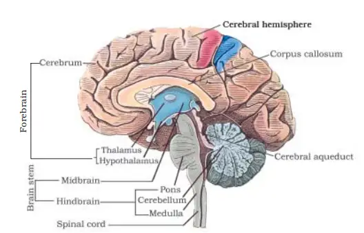
The human brain has following parts
Forebrain
Midbrain
Hindbrain
Forbrain
It consist of cerebrun, thalamus and hypothalamus Cerebrum:-
A deep cleft called longitudinal fissure divides the brain/ cerebrum into two halves i.e., cerebral hemispheres.
The two cerebral hemispheres are joined together by bundles of densely packed nerve fibres, called corpus, callosum.
The outer surface of cerebrum, the cerebral cortex, is called grey matter due to grayish appearance; the cell bodies of the neurons are concentrated in this region. It contains motor areas, sensory areas & association areas.
Inner to the cortex is the white matter that contains myelinated nerve fibres in the form of nerve fibre trad.
Thalamus:-
The cerebrum wraps around a structure called thalamus which is the major coordinating centre for sensory and motor signalling.
Hypothalamus:-
Hypothalamus lies at the base of thalamus, that contain a number of centres with control body temperature, urge for eating and drinking. It also contains several groups of neurosecretory cells which secrete hypothalamic hormones
Midbrain:
It is located between thalamus/ hypothalamus of the fore brain and the pons of hindbrain.
The dorsal portion of it consists of four round swelling called corpora quadrigemina.
Hindbrain:
pons control respiratory activity.
Cerebellum control balance and posture of the body
Medulla oblongata controls involuntary actions.
Brain Stem:
It forms the connection between the brain and spinal cord. Three major regions make up the brain stems are midbrain, pons and medulla oblongata.
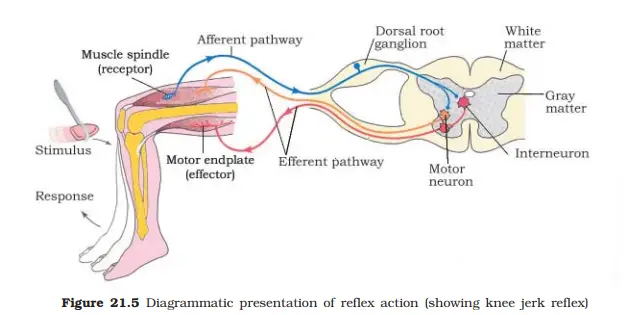
Reflex action and Reflex arc:
The entire process of response to a peripheral nervous system stimulation, that occurs involuntarily, that is, without conscious effort or thought and requires the involvement of a part of the central nervous system is called a reflex action.
The reflex pathway comprises at least one of afferent neuron (receptor) and one efferent neuron (receptor) appropriately arranged in a series. Thus, the coordination series of stimulus and response from reflex arc.
This portin has been deleted from CBSE Syllabus
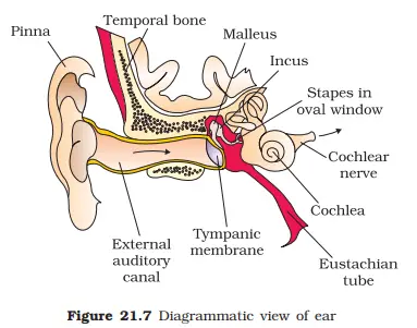
HUMAN EAR
The ear performs two sensory functions:-
Hearing
Maintenance of body balance
Structure of ear:-
Ear consists of three parts ie. External, middle & internal ear External Ear:-
The external ear consists of pinna and external auditory meatus (auditory canal). The external auditory canal reads inwards & extends up to the tympanic membrane (ear- drum) which separates the middle ear from external ear. Middle Ear:-
The middle ear is an air field chamber, which is connected to pharynx by Eustachian tube.
The middle ear contains three ossicles called malleus, incus, (Anvil), & stapes. The middle ear communicates with the internal ear through the oval window & round window.
Internal Ear:-
The inner ear is a fluid filled chamber called labyrinth, which has two parts, an outer bony labyrinth, inside which a membranous labyrinth is floating in the perilymph. The membranous labyrinth is filled with fluid called endolymph.
The labyrinth is divided into two parts, the cochlea & vestibular apparatus.
Cochlea is the coiled portion of the labyrinth. At the base of the cochlea, Scala vestibuli ends at the oval window, while the scala tympani terminates at the round window that opens to the middle ear.
ORGAN OF CORTI:-
The Organ of corti is the structural unit of hearing. It consists of hair cells that act as auditory receptors. VESTIBULAR APPARATUS:-
It is composed of three semi – circular canals & otolith, which has two parts namely the utricle & saccule.The crista and macula are the specific receptors of the vestibular apparatus responsible for maintenance of balance of the body and posture MECHANISM OF HEARING:-
The pinna collects the sound waves and directs them to the eardrum.
The eardrum vibrates in response to the sound waves and these vibrations are transmitted through the ear ossicles (malleus, incus and stapes) to oval window through ear ossicles & from these they reach the fluid of the cochlea where they generate waves in the perilymph & endolymph.
The waves in the lymph induce a ripple in the basilar membrane.
These movements of the basilar membrane, bend the hair cells of the organ of corti which press them against the tectorial membrane. This pressing generates nerve impulses in the associated afferent neuron.
The afferent fibre transmits the impulse via auditory nerve to the auditory cortex of the cerebrum (Brain) where the impulses are analyzed & the sound is recognized.
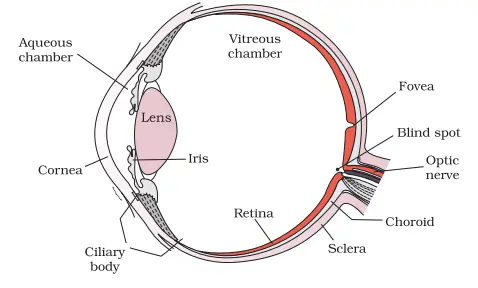
HUMAN EYE
The organ of sight is a pair of eyes in humans.
Our paired eyes are located in the socket of the skull called orbits.
The adult human eyeball is nearly spherical in structure & consists of three concentric layers, the outermost sclera, middle choroid and innermost retina.
Sclera is an opaque outermost covering which in the front forms the transparent cornea.
The middle choroid is highly vascular ,contains many blood vessels and pigmented that prevents internally reflected light within the eye; just behind the junction between cornea & sclera the choroid becomes thicker forming the ciliary body.
The ciliary body itself continued forward to form a pigmented and opaque structure called iris and it controls the dilation or constriction of pupil.
The eye all contains a transparent crystalline structure called lens which is holded by the ciliary body and in front of less the aperture surrounded by iris is called pupil.
The interior chamber of the eye is filled with aqueous humor & the posterior chamber has a gelatinous material called vitreous humor.
The inner layer is the retina & it contains three layers of cells from inside to outside i.e., ganglion cells, bipolar cells & photoreceptor cells. The photoreceptor layer contains rods and cones, which contains light sensitive proteins called photopigments.
The daylight vision (photopic) and colour vision (red, green and blue light) are functions of cones and the twilight vision (scotopic) is the function of rods (contain purplish red protein called rhodopsin, which contain derivatives of Vitamin A).
Photoreceptor cells are not present in that region where optic nerve leaves the eye & the retinal blood vessels enter the eye is called blind spot.
Lateral to blind spot there is a yellowish pigmented spot called macula lutea with a central pit called fovea.
Fovea is the region where only cones are densely packed & it is the point where acuity (resolution) vision is the greatest.
MECHANISM OF VISION:- (Binocular vision):-
Retina receives light rays through the cornea and lens generates impulses in the rods & cones.
The photosensitive compounds in the human eye are composed of opsin & retinal (Aldehyde or derivative of vitamin A).
The received light induces dissociation of retinal from opsin which causes the change in the conformation of opsin which caused change in the permeability of membrane; as a result potential difference generated in photoreceptor cells.
They are transmitted via bipolar neuron to ganglion cells; the axons of ganglion cells bundle up to form optic nerve which takes the impulse to the visual cortex of the brain.
In brain neural impulses are analyzed & image formed on the retina is recognized.