Protoderm:- It is the outermost portion of the primary meristem found at the apex of stem and root. It develops into epidermis.
Procambium:- It develops into primary vascular tissues. It forms the isolated strands of elongated cells, very near to the central region.
Ground Meristem:- It develops into ground tissues in the later stage of growth, they become differentiated into hypodermis, cortex, Endodermis, pericycle, medullary rays & pith.
Classification based on origin:-
Primary Meristem:- These meristems are derived during the early embryonic stage. They divide rapidly & differentiated into primary permanent tissues. They are formed in the growing apical region of root & shoot.
Secondary Meristem:- These meristems appear in later stages of development. They lie lateral in position in both stems & roots. Examples of secondary meristems are: cork cambium & interfascicular cambium. They allow secondary growth in tissues.
The Xylem formed in the primary plant body by procambium is known as primary xylem.
The first or earlier formed primary xylem is called protoxylem.
The later formed primary xylem is known as metaxylem.
In stems protoxylem lies towards the pith and metaxylem lies towards the periphery; such an arrangement of xylem is called endarch.
In roots, protoxylem lies towards the periphery (cortex), while metaxylem lies towards the pith (centre), such an arrangement of xylem is called exarch.
[The xylem elements formed later by fascicular and interfascicular cambia constitute secondary xylem.]
TISSUE SYSTEMS IN PLANTS
There are three tissue systems in plants based on the structure and location in the plant body. They are epidermal tissue system, vascular tissue system and ground or fundamental tissue system.
EPIDERMAL TISSUE SYSTEM
It consists of the epidermal cells, which forms the epidermis, epidermal appendages (like root hair & trichomes) & stomatal apparatus.
Epidermis is usually single layered. Epidermal Cells are parenchymatous with a small amount of Cytoplasm lining the cell – wall & large vacuole.
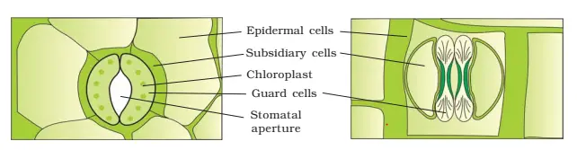
Stomata are the structure present in the epidermis of the leaves. It regulates the process of transpiration & gaseous exchange. Each stoma (stomata) is composed of two bean shaped cells called guard cells which encloses stomatal pore. The outer walls of guard cells are thin & inner walls are highly thick.
In some species guard cells are surrounded by subsidiary or accessory cells. In monocots, the guard cells are dump – bell shaped.
(a) stomata with bean-shaped guard cells (b) stomata with dumb-bell shaped guard cell
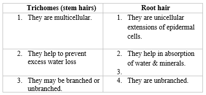
EPIDERMAL APPENDAGES:-
GROUND TISSUE SYSTEM
All tissues, except epidermis and vascular bundles constitute the ground tissue system. Its various components are hypodermis, cortex, endodermis, pericycle, pith, medullary rays and pith.
It consists of simple permanent tissues like parenchyma, collenchyma & sclerenchyma.
VASCULAR TISSUE SYSTEM:-
It Includes Xylem & phloem which occurs as discrete strands called vascular bundles.
On the basis of arrangement of xylem & phloem in the vascular bundle, there are 3 types of bundles:-
1.RADIAL:
The xylem & phloem alternate with each other separated by parenchymatous cells. It is found mainly in roots.
2.CONJOINT:-
The xylem & phloem are present together in the same bundle on the same radius. They are of 2 types:- (i)Collateral:-
The xylem & phloem lie together on the same radius. The xylem lies inwards & phloem outwards. (a)OPEN VASCULAR:-
In this the cambium is found to be present in b/w the xylem & phloem. They are found in dicot stems. For example:- Helianthus (sunflower). CLOSED:-
When the cambium is absent, the vascular bundle is called a closed bundle. For example:- Zea mays (Maize). (ii) Bicollateral
In this 2 groups or patches of phloem one on each side of the centrally located xylem is present. (iii) CONCENTRIC:-
A vascular bundle in which one tissue is completely surrounded by the other is called concentric.
Amphivasal:- Phloem lies in the centre & completely surrounded by xylem.
Ambhiribal:- Xylem lies in the centre & completely surrounded by phloem.
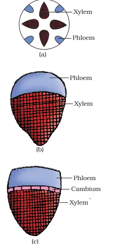
Various types of vascular bundles :(a) radial (b) conjoint closed (c) conjoint open
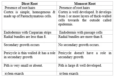
Differentiate between dicot root and monocot root
Identification features of dicot root:-
Presence of root hairs.
Endodermis with Casparian strips.
Absence of pith.
Radial bundle less than 8.
Presence of exarch xylem.
Identification features of Monocot root:-
Presence of root hairs.
Endodermis with passage cells.
Presence of pith.
Radial bundles more than 8.
Xylem exarch.
Presence of epidermis.
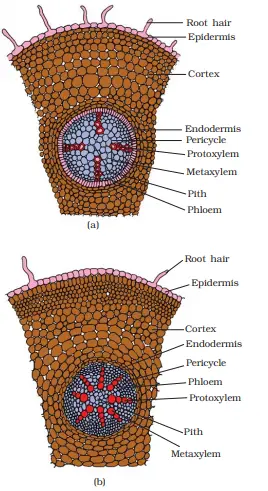
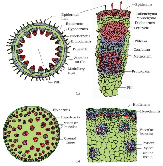
(a) Dicot root (Primary) : (b) Monocot root
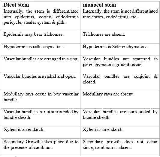
Differentiate between Dicot stem and monocot stem
(a) Dicot stem (b) Monocot stem
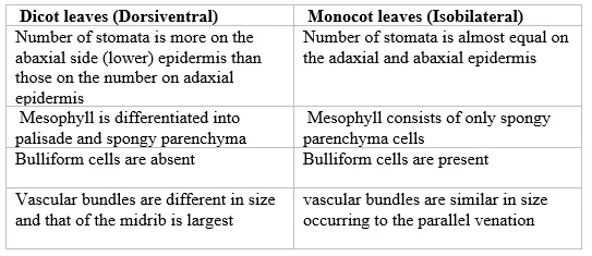
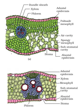
Differentiate between dicot and monocot leaves
(a) Dicot leaf (b) Monocot Leaf
This portion is excluded from cbse syllabus
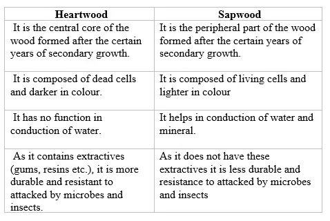
Differentiate between heartwood and sapwood
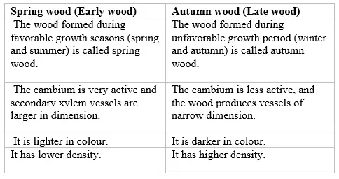
differentiate between springwood and autumn wood
Secondary Growth
Most dicotyledonous plants exhibit an ability to increase in girth, this increase is called secondary growth.
The tissue involved in secondary growth are two lateral meristems (secondary meristem) i.e.. Vascular cambium & cork cambium. Secondary Growth in DICOT STEM:-
It is achieved by fascicular, inter- fascicular & cork cambium, i.e., the lateral meristem.
Certain fascicular cambium and inter fascicular cambium join to form a complete ring called cambial ring.
The cells of the cambial ring undergo mitotic division and produce secondary phloem on its outer side & secondary xylem on its inner side.
A long certain radius, secondary medullary rays are also formed by the cambium.
With the increase in the girth/ diameter of the stem, a secondary protective tissue replaces the epidermis which becomes broken.
It is formed by cork cambium (Phellogen) which produces cork (Phellem) on its outer surface & secondary cortex (Phelloderm) on its inner side.
The phelloderm, phellogen & phellem together constitute periderm.
The trunk becomes thick to support the canopy.
Secondary growth in Dicot roots
In the dicot root, the vascular cambium is completely secondary.
In the dicot root, vascular cambium arises as some cells of conjunctive tissues.
On the inner edge of the phloem and the cells of the pericycle lying opposite to the xylem tissue become meristematic.
These patches of meristematic cells join and form a cambial ring which is initially wavy in outline but later becomes circular.
Further events are similar to that of Dicot stem
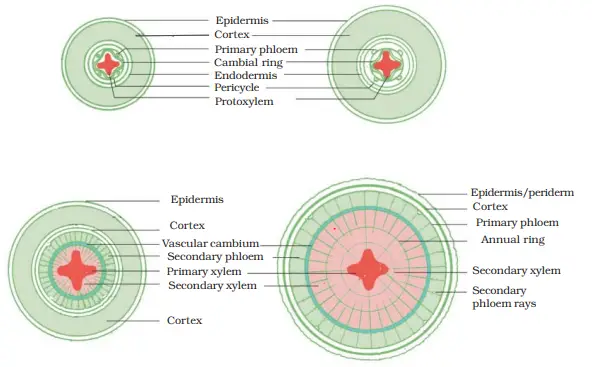
Different stages of the secondary growth in a typical dicot root What is the significance of secondary growth?
It replaces old, non- functional tissues.
Commercial cork is a product of secondary growth.
It provides fire proof, insects proof & insulted cover around the older plant part.
Wood is the product of secondary growth.
Cork has suberin deposition which makes it impervious to water.