Streaming of cytoplasm in unicellular organisms is the simplest form of movement. Animals move their body parts like limbs, jaws, tentacles etc.
Animal’s cells also show movement of cilia & flagella. In paramoecium cilia helps in movement of food into cytopharynx & also for locomotion.
Hydra uses the tentacles and human beings uses the limbs to change the posture of the body as well as for locomotion.
TYPES OF MOVEMENTS
Cells of Human Body Show four basic types of movements:-
Amoeboid Movement – Movement with the help of Pseudopodia. Macrophages & leucocytes exhibited amoeboid movement.
Ciliary Movement – Movement with the help of cili .Ciliary Movement occurs in most of our internal tubular organs that are lined by ciliated epithelium. For ex:- removal of dust particles & foreign substances through the trachea, passage, of ova through female reproductive tract.
Flagellar Movement- flagellar movement is involved streaming of Spermatozoa in the female reproductive tract of humans.
The above three movements are called non- muscular movement:- iv. Muscular Movement- Move with the help of muscles is called muscular movement. Move of jaws, limbs, eyelids etc. are Muscular movement involving striated muscles. Movement of food in the alimentary canal and movement of urine in the ureter are movement involving smooth muscles.
MUSCLES
Muscle is a specialized tissue that arise from mesoderm & are made up of muscle fibres. Special Properties:-
Contractibility:- The cells of muscles can be shortened considerably & return to the original relaxed state.
Excitability:- It is due to the energy stored in electric potential difference across the plasma membrane.
CLASSIFICATION OF MUSCLES
Muscles have been classified using criteria like location, appearance & nature of regulation of their activities.
In humans, the muscles are broadly classified into 3 categories as given below:-
Skeletal Muscles
(Striated or Striped)
Smooth Muscles
(Visceral or Unstriated)
Cardiac Muscles
Striation are prominent.
Striation are absent.
Striation are faint.
Voluntary in function.
Involuntary
Involuntary
Cells are multinucleated.
Uninucleated
Uninucleated
Fibres are cylindrical & unbranched. (controlled)
Fibres are spindle shape and unbranched
Fibres are cylindrical, branched & show intercalated disc.
Controlled by voluntary nervous system.
Controlled by Autonomous Nervous system.
Controlled by autonomous nervous system (not direct)
Ex- Muscles of limbs.
Ex- Muscles of visceral organs (internal organ)
Ex- Heart Muscles
Skeletal Muscles
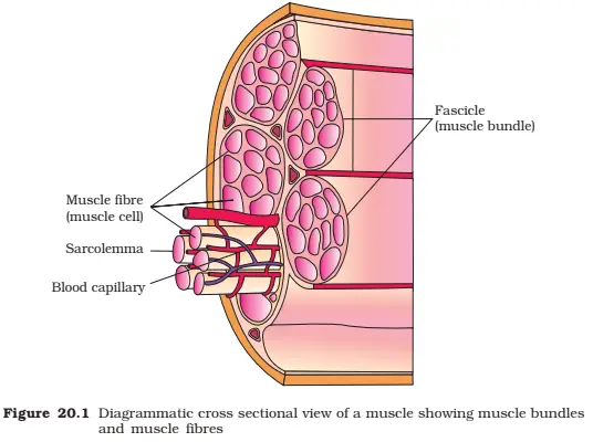
Skeletal muscle is made up of no. of muscle bundles or fascicles held together by fascia (connective tissue).
Each muscle bundle has a no. of muscle fibres or cells with a membrane called sarcolemma that encloses the cytoplasm called sarcoplasm.
The sarcoplasm contains mitochondria (sarcosomes), Endoplasmic Reticulum (sarcoplasmic Reticulum) and many thin parallel arranged filamentous structure called myofibrils or myofilaments.
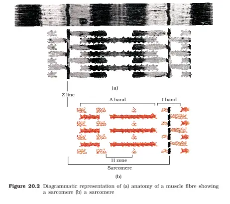
Each myofibril has throughout its length, alternate dark & light bands that gives the striated appearance of the muscles.
The light band contains actin & are called I- band (Isotropic band).
The dark band contain myosin & are called A – band (Anisotropic band).
In the centre of the I- band is an elastic fibre called Z – line to which the actin filaments are firmly attached.
The portion of the myofibril b/w two Z – lines is a sarcomere, the functional contractile unit.
The central part of the A – band not overlapped by the thin filament is called H – zone.
STRUCTURE OF CONTRACTILE PROTEIN:
Actin Filament (thin filament, I-band)
It contains 3 proteins namely actin, tropomyosin & troponin.
Each actin filament consists of 2 F-actin (filamentous) that are helically bound to each other. It is a polymer of G – actin (Globular).
Two filaments of Tropomyosin also run close to the F- actin along their length.
Troponin is a complex protein found at regular intervals on the tropomyosin.
In resting state a sub – unit of troponin mask the active binding site of myosin.
Myosin Filament:- (Thick filament, A- band)
It is a polymer of meromyosin. Each myosin has 2 important parts, a globular head with short arm & a tail.
The head with short arm is called heavy meromyosin (HMM) & the tail is called light meromyosin (LMM).
The HMM component projects outward at regular distance at an angle from the surface of the polymeric meromyosin, it is known as cross – arm.
The globular head, functions as ATPase enzyme & has binding sites for ATP & active site for actin.
Mechanism of Muscles Contraction
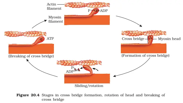
The contraction of muscles is best explained by the sliding filament theory. This theory states that contraction of muscle fibre is due to the sliding of the thin filament (Actin) & thick filament (myosin).
Muscle contraction is initiated by a neural signal from the CNS through a motor neuron.
When the neural signal reaches the neuro mensular junction, it releases a neurotransmitter i.e., Acetylcholine; which generates an action potential in the sarcolemma. This spread through the muscle fibre and causes release of calcium ions from the sarcoplasmic reticulum into the Sarcoplasm.
The Ca++ binds to the sub – unit of troponin & brings about conformational changes; this removes the masking of active site for myosin.
The myosin head binds to the active site on actin to form a Cross Bridge This utilizes energy from the hydrolysis of ATP. This pulls the actin filament towards the centre of A- band. As a result the Z- line also pulled inwards causing shortening of Sarcomere i.e., Contraction of muscles.
Hence, during muscle contraction, the length of A – band remains unchanged while that of I – band decreases.
Muscle Relaxation
The myosin goes back to its relaxed state, a new ATP binds & the cross- bridge is broken & the actin filament slide out of A- band.
The cycle of cross bridge formation & cross bridge breakage continues till the calcium ions are pumped back into the sarcoplasmic reticulum which leads to blocking the active site on actin filament & Z – line returns to original position i.e., Relaxation.
Types of Skeletal Muscles
Based on amount of red – coloured pigment myoglobin present in them, skeletal muscles are of two types:-
Red Muscle Fibre
White Muscle Fibre
Red Muscle Fibre
They are dark red muscle fibre due to the presence of abundant myoglobin in them.
They have more no. of mitochondria.
Sarcoplasmic reticulum is less.
They depend on aerobic process for energy.
These muscle fibres are thinner.
For example:- Extensor muscle of human back.
White Muscle Fibre
They are pale or whitish as they have less myoglobin.
They have less no. of mitochondria.
Sarcoplasmic reticulum is more.
They depend on anaerobic process for energy.
These muscle fibres are thicker.
For example:- eyeball muscles.
Disorders of Muscular system
Muscular Dystrophy:- It is a genetic disorder resulting in progressive degeneration of skeletal muscles.
Myasthenia Gravis:- It is an autoimmune disorder affecting the neuromuscular junction leading to progressive weakening & paralysis of skeletal muscles.
Tetany:- It refers to the continued state of contraction or wild – contraction of muscles due to low calcium ions in body fluid.
Skeletal system
It consists of a framework of bones & cartilages. functions
It plays a vital role in movement & locomotion.
It provides protection to many vital organs.
It serves as a reservoir of calcium & phosphates.
The bone marrow of long bones produces blood cells (Haemopoiesis).
Division of Skeleton
Human body is made up of 270 bones which are used variously to form 206 bones.
Axial skeleton:
It comprises of 80 bones which includes skull, vertebral column, ribs and sternum. Skull bones:
There are 8 flat bones in the cranium. They are articulated immovably with the help of fibrous connective tissues forming sutures.
The facial region has 14 skeletal elements. A single U- shaped bone called hyoid bone is present at the base of buccal cavity.
Each middle ear has three small bones called ear ossicles. They are malleus, incus and Stapes.
Human skull is dicondylic, the skull region articulate with vertebral column with the help of two occipital condyle.
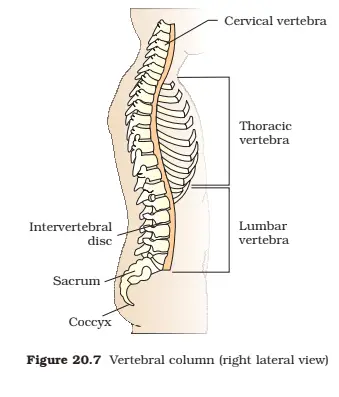
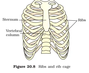
Vertebral column:
It is formed of a series of 26 bones called vertebrae.
It includes 7 cervical vertebrae in the neck region. the first cervical vertebrae is Atlas, which articulate with the occipital condyle of the skull.
There are 12 thoracic vertebrae and 5 lumbar vertebrae and the lowermost part of the vertebral column is formed of two fused bones, the sacrum and coccyx ( coccygeal).
Functions of vertebral column:
Provide protection to the spinal cord.
Support the head.
Allow flexibility and bending of the back of the body.
Also serve as the point of attachment for the ribs.
Sternum :- (chest bone)
It is a flat, dagger shaped bone located on the ventral middle of thorax.
Ribs:-
There are 12 pairs of ribs in an adult human being.
Each rib is a thin flat bone articulates dorsally with the respective thoracic vertebrae & ventrally with the sternum.
It has two articulation surfaces on its dorsal end & is called Bicephalic.
True Ribs:-
The first 7 pairs of ribs are attached directly to the sternum with the help of hyaline cartilage & are called true ribs. False Ribs:-
The 8th, 9th & 10th pair of ribs joined the 7th rib with the help of hyaline cartilage & does not articulate directly with sternum. Hence, called vertebrochondral or false ribs. Floating Ribs:-
The 11th & 12th pair of ribs remains free anteriorly & are called floating ribs. FUNCTIONS OF RIBS
They product the heart, large blood vessels & lungs.
Bear respiratory muscles.
Lower 2 pairs of ribs protect the kidney.
Appendicular Skeleton:-
It comprises of 126 bones which include pectoral & pelvic girdles & limb bones of arms & legs. Pectoral Girdle (Shoulders)
It consists of two bones i.e., Scapula and Clavicle (collar bone).
Scapula is a large triangular flat bone which consists of sharp ridge i.e., Spine. The end of spine projects as a flattened & expanded portion called Acromion which articulates with the clavicle.
Below the acromion there is a depression called Glenoid cavity which articulates with the head of humerus.
Clavicle is a long, slender bone with two curvatures.
Functions of Pectoral Girdle:-
It provides an attachment point for numerous muscles that allow shoulder & joints to move.
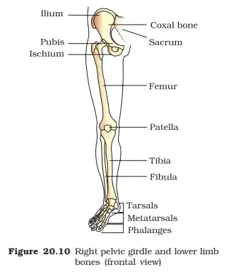
Pelvic Girdle (hips):-
It consists of two coxal bones which is formed by the fusion of 3 bones i.e., ilium, ischium & pubis.
At the point of fusion of 3 bones a cavity called Acetabulum is present where head of femur articulates.
Functions of Pelvic Girdle:-
It provides articulation to the bones of the legs, supports & protects abdominal viscera (limbs).
Bones of Limbs:- Forelimb:-
Each arm consists of 30 bones. The upper arm has one bone humerus while the lower arm is supported by radius & ulna, which together makes up the elbow.
The wrist has 8 carpals & the palm has 5 meta carpals.
The thumb has 2 phalanges, while the other fingers have 3 phalanges (small bones) each.
Hind Limb:-
Each hind limb has 30 bones.
The thigh is supported by longest & heaviest bone of the body called femur.
Tibia and fibula together support the shank of the leg.
There are 7 tarsals in the ankle.
The bones of the foot are called metatarsals, which articulates with the tarsal at one end and with phalanges at the distal end.
The big toe has two phalanges & the other toes have 3 phalanges each.
A cup shaped bone called patella (knee cap) covers the knee ventrally.
JOINTS:-
A joint is a place of articulation between two or more bones or between a bone and cartilage.
The force generated by the contraction of muscles is used to carry out movements through joints which acts as a fulcrum.
Depending on the extent of mobility, the joints are classified into three types:-
Fibrous joints (Immovable)
Cartilaginous joints (slightly movable joints)
Synovial joints (freely movable joints)
Fibrous Joints:-
In these joints the articulating bones are very tightly held with the help of fibrous connective tissue in the form of sutures.
For ex.- Sutures between the skull bones, articulation of roots of teeth with the sockets of jaw bones etc. Cartilagenous Joints:-
In such joints the articulation of bones allows very little movement. The opposing surface of the bones are connected by fibrocartilage.
For ex- Joints between adjacent vertebrae in the vertebral column. Synovial Joints:-
This type of joints allow extensive movement of articulating bones upon each other & characterized by the presence of fluid filled cavity between articulating bones is known as synovial fluid, which reduces the friction betwen the bones during movement.
There are 5 different types of synovial joints:-
Ball & Socket Joints:- Ex- between humerus & pectoral girdle, between femur & pelvic girdle.
Hinge Joints:- Ex – knee joint & elbow joint.
Pivot Joints:- Ex- between atlas & occipital condyles.
Gliding Joints:- Ex- between the Carpals, and between the tarsals.
Saddle Joints:- Ex- between carpals & metacarpals of thumb.
DISORDERS OF BONES:-
Arthritis:- It is the inflammation of joints.
Osteoporosis:- It is an age dependent disorder of the bones, characterized by low bone mass, deterioration of micro architecture of bone & increased fragility.
Gout:- It is the inflammation of joints due to excess accumulation of uric acid.