Study of different structures found within the cell and of the fundamental processes related to plant, animal and bacterial cell is called cell biology.
Cell membrane
These are semi-permeable i.e they allow passage of some solute through them but not the other. It is made up of proteins and lipids. Lipids are phospholipids, form a fluid in it (Fluid mosaic model).
Different types of proteins are there i.e integral protein, Peripheral protein and train-membrane protein. Some penetrates only a part of lipid bilayer while the other penetrates all the way through usually the hydrophobic portion of protein interacts with lipids. The hydrophilic portion face the aqueous content at the membrane surface. Sometimes protein form channel or pores which span the thickness of membrane allowing passage of solute.
Some membrane protein function as enzyme/receptors for legends /electron carrier. Some proteins are glycosylated (glycoprotein) and help in cell to cell adhesion some protein play an important role in immune response.
Junctions of Cell Membrane
(i) Separates the interior of the cell from the exterior and also control exchange of gases, solute and maintain osmotic balance.
(ii) Bound the cell organelles for example -mitochondria, Golgi body.
(iii) Act as site for photosynthesis or oxidative phosphorylation.
Cell organelles
An organelle is a cellular structure with specialized function that forms a part of the cell. Some of the important organelles are
(a) Nucleus
(b) Cytoplasm
(c) Endoplasmic reticulum
(d) Ribosomes
(e) Golgi apparatus
(f) Lysosomes
(g) Mitochondria
(h) Plastids
(i) Cytoskeleton
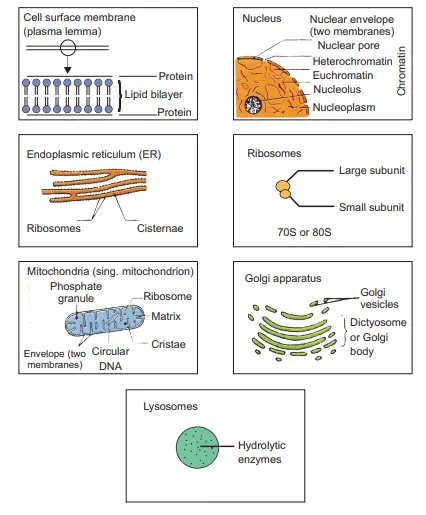
Nucleus
Largest cell organelle. Spherical, ovoid in shape about 10nm in diameter bounded by nuclear membrane perforated by nuclear pores, meant for exchange of substance between nucleus and cytoplasm. Outer membrane is in continuation with membrane of ER membrane. Nucleus contain chromatin is composed of DNA and histone protein. Both of them are organized into a bead like structure called nucleosomes.
During cell division chromatin get condensed and take shape of chromosome.
Certain areas in chromatin which are tightly coiled and darkly stained transitionally inactive called heterochromatin whereas loosely coiled chromatin with slightly strained and transcriptionally active is called euchromatin.
Euchromatin contains a greater number of active genes than heterochromatin. Nucleolus a rounded structure within the nucleus contain large amount of DNA and RNA. It synthesize ribosomal RNA which assemble with protein to form ribosomes.
Cytosol (Cytoplasm)
It is the brown, aqueous substance inside the cell which contain cell organelles and insoluble cellular waste/storage product.
Composition –
90% water, dissolved ion, salts, sugar, amino acid, fatty acid, nucleotides vitamins, dissolved gases, microfilaments.
Note :-
Protoplasm – Nucleus + cytoplasm
Cytosol – Soluble part of cytoplasm
Cytoplasm store vital chemical and act as a site for metabolic pathways such as glycolysis.
Endoplasmic reticulum (Intra cellular transport channel of the cell)
Complex network of membrane throughout the cytoplasm. It consist of flattened membrane bound sac called cisternae.
Rough ER
Smooth ER
ER covered with ribosomes
ER without attached
It is lite of protein Synthesis
It is a site of lipid synthesis
ER help in transportation of protein through its cisternae for example – transportation of protein through Golgi apparatus.
Non-membranous organelle (Ribosomes)
It is site of protein synthesis. It consists of two sub units one small and one large.
Based on their sedimentation rate in centrifuge machine. These are of two types
-70s (5+30s) -: found in bacterial cell, mitochondria.
-80S (60s, 40) found in plant cell and animal cell
Ribosomes are chemically ribonucleoprotein i.e. they are made up of RNA and protein.
Polysomes-
When number of ribosomes get attached to mRNA during translation.
Golgi apparatus
It consist of pile of flattened membrane bound sac called cisternae. cisternae are formed by fusion of vesicles which bud off from ER and this fusion takes place on the outer convex surface of Golgi apparatus.
It synthesize lysosomes and it is packaging and dispatching unit of cell. Addition carbohydrate moiety to the protein by a process of glycosylation.
Lysosomes
Small single cell bound organelle containing hydrolytic enzyme such as proteases, nucleases and lipases.
These enzymes are synthesized on ER and transported via cisternae to the Golgi apparatus from which they are pinched off as vesicles and form primary Lysosomes.
Now primary lysosomes and phagocyte vesicle form phagolysosomes which digest the material with the help of hydrolytic enzymes which have phagocytosed by the cell.
Mitochondria
Elongated organelle bounded by two membrane inner membrane is folded to form cristae. Matrix of mitochondria contain 70s ribosome, DNA and phosphate granules. It is a site of Krebs’s cycle. Mitochondria is called poorhouse of cell as cristae site of oxidative phosphorylation and electron transfer due to which ATP is formed.
Plastid
Present only in plant cell and bounded by cell membrane. Chloroplast contain chlorophyll, carotenoid and carry out photosynthesis. The brown substance of chloroplast is called stroma in which flattened fluid filled sacs called thylakoids are present two grana are connected.
Membrane of thylakoids are filled with enzymes and electron carrier.
Chromoplast – Non-photosynthetic colored plastid containing red, yellow and orange pigment. These are present in the cell of fruit and flower and impart colors to them and help in pollination to attract insects/
Leucoplasts – Colorless plastid lacking pigment act as food storage sight and are of three types –
amyloplasts (Store starch) ,lipidoplast (stores lipid), proteotoplasts (proteins).
Cytoskeleton
The living cells must maintain their shapes. A network of fibrous protein structure carries out this task known as cytoskeleton. It is of two types :-
i) Microtubules -: these are unbranched, hollow, cylindrical organelles which consist of protein called tumbling. In spindles, cilia and flagella microtubules undergo sliding motion, these are responsible for movement of chromosome, flagella, cilia and Golgi vesicles etc.
ii) Microfilaments – Made up of actin protein and occur in sheath or bundle just below the cell membrane. These are involved in endocytosis, exocytosis and movement of cell.
Tissues
A group of cells similar in structure that work together to perform a particular function forms a tissue.
All types of tissues have two basic components :
(a) Cells having common origin and function
(b) Intercellular substances : Are nonliving, fibrous, jelly-like substances.
Importance of tissues :
1. Formation of tissues has brought about divisor. of labor in multicellular organisms.
2. Tissue become organized to form organs and organs into organ systems.
3. Workload of individual cell has decreased due to origin of tissues.
4. As a result of improved organization and higher efficiency, multicellular organisms have higher survival.
Classification of tissues :
Tissues are broadly classified as animal tissues and plant tissues.
Plant Tissues
Plant tissues can be broadly divided into two main types. These are Meristematic tissue and permanent tissue.
Meristematic Tissues :
A meristematic tissue constitutes a group of actively cells present in the growing region of plant, e.g., the tips of roots and stems.
These tissues are responsible for increasing the length and girth of the plant.
Characteristics of meristematic tissues :
The cells of the meristematic tissues are similar in structure and have thin cellulose cell walls.
The cells may be spherical, oval, polygonal or rectangular in shape.
The cells have dense protoplasm with prominent nuclei.
Vacuoles in these cells are either small or absent.
On the basis of their position in the plant body, meristematic tissues are classified into three types :- Apical, Lateral and Intercalary.
Apical meristems :
These are present at the tips of roots, shoots, branches and leaves.
It brings about the elongation of the root and stem. It results in increase in the Hight of the plant, which is called primary growth.
Lateral meristems :
These are present along the lateral side of the stems and roots. For example : cork cambium.
It causes the organ (stem or root) to increase in diameter and girth. This is called secondary growth.
Intercalary meristems :
They are located at the base of leaves or internodes, e.g., stems of grasses and other monocots and below the nodes (e.g. mint).
It produces an increase of length of organ such as leaves and internodes.
Functions : Meristematic tissue acts as a parent tissue from which other tissues develop.
These tissues take part in growth by formation of new cells.
The place of injury in plants is healed up by the formation of new cells by meristems.
Permanent tissues
A permanent tissue is a group of cells, which is derived from the meristematic tissues, but these cells have lost the power of division temporally or permanently.
Note : The development process by which cells which has been derived from meristematic tissues take a permanent shape, size and function is called differentiation.
Permanent tissues are of two types :- simple and complex permanent tissue.
Simple permanent tissues :
These tissues are composed of cells which are structurally and functionally similar. These tissues are of three types parenchyma, collenchyma and sclerenchyma.
Parenchyma :
Parenchyma forms the bulk of plant body. It consists of thin walled living cells.
The cells are isodiametric, i.e., equally expanded on all sides.
The cell wall is thin and encloses a dense cytoplasm which contains a small nucleus and a large central vacuole.
The intercellular spaces are abundant.
The parenchyma is present in all the organs of the plants, i.e., roots, stems, leaves, flowers, fruit and seeds.
Function :
The main function of parenchymatous tissue is storage of food, e.g., starch in the parenchyma of cortex of potato tuber.
Parenchyma forms the framework of all the plant organs and tissues like cortex. Pith etc.
Parenchyma serves as packing tissue to fill the spaces between other tissues.
It stores waste materials of plants such as gum, crystals etc.
The intercellular air spaces of parenchyma cells allow gaseous exchange.
If chloroplast is present, the parenchyma tissue is called chlorenchyma and it perfoms photosynthesis.
In aquatic plants, large air cavities are present in parenchyma to give buoyancy to the plants to help then float. Such a parenchyma type is called aerenchyma.
Collenchyma :
Collenchyma is usually found in 3-4 layers beneath epidermis in stem, petioles and leaves of herbaceous dicot plants.
The cells of this tissue are living, elongated and irregularly thickened at the corner.
In collenchyma, intercellular spaces are generally absent.
Functions :
It provides the mechanical support, protection, flexibility and elasticity to the plant’s organs.
It allows easy bending in various parts of the plant (leaf, stem) without breaking.
When cells of collenchyma contain some chloroplasts, they manufacture sugar and starch.
Sclerenchyma:
Sclerenchyma cells are dead cells and they are devoid of protoplasm.
They are long and narrow as the walls are thickened due to lignin, such cell walls are called lignified.
The cells of sclerenchyma are closely packed without intercellular spaces.
Cells of sclerenchyma are of two types : fibres and sclereids.
Fibres consist of very long, narrow, thick and lignified cells. Sclereids are irregular shaped.
This tissue is present in stems, around vascular bundles, in the veins of leaves and in the hard covering of seeds and nuts. Husk of coconut is made of sclerenchymatous tissue.
Functions :
The sclerenchyma is mainly mechanical and protective in function.
It gives strength, rigidity, flexibility and elasticity to the plant body and, thus, enables. It to withstand various strains.
Complex permanent Tissues
The complex tissue consists of more than one type of cell having a common origin. All these cells coordinate to perform a common function.
Complex tissues are of two types : Xylem or wood and phloem or bast.
Xylem and phloem are both conducting tissues and also known as vascular tissues; together both them constitute vascular bundles.
Xylem
Xylem is a vascular and mechanical tissue.
Xylem is composed of cells of four different types:
Except xylem parenchyma, all other elements are dead and bounded by thick lignified wall.
Tracheid’s and vessels are tubular structures.
Functions :
The main function of xylem is to carry water and mineral salts upward from the root to different parts of shoots, hence also called water conducting tissue.
Since walls of tracheids, vessels and Sclerenchyma of xylem are lignified, they give mechanical strength to the plant body.
The parenchyma stores food and helps in the sideway conduction of water.
Phloem
Phloem (bast) is a living conducting tissue. It also contains tubes just like xylem but does not perform mechanical function.
Phloem is composed of following four elements or cells :
Sieve tubes are slender, tube like structures with perforated walls.
Companion cells are living parenchymarous cells lying on the sides of the sieve tubes. Sieve tube and companion cells have close cytoplasmic connection with each other through fine pits.
Phloem fibres are thick walled fibres with simple pits.
Phloem parenchyma are thin walled, living cells of parenchyma of phloem.
Function :
Phloem transports (conducts) photosynthetically prepared food materials from the leaves to the storage organs and later from storage organs to the growing regions of the plant body.
Protective tissues
Protective tissues are a part of plant tissue system. Protective tissues include epidermis and cork.
Epidermis
It is the outermost protective layer of plant organs.
The epidermis is usually made of a single layer of cells.
Cells of epidermis are elongated and flattened, without intercellular space. They are living cells but their inner contents are similar to parenchyma cells.
In leaves, epidermis bears small pores called stomata.
In some plants living in very dry habitats, the epidermis may be thicker since protection against water loss is critical.
Functions :
The function of epidermis is the protection of plant from injury and infection.
Cuticle of epidermis also helps to reduce water loss by evaporation to prevent dessication.
Stomata present in the epidermis allow gaseous exchange to occur during photosynthesis and respiration.
It also facilitates transpiration.
Cork (or phellem) :
Cork cells are dead cells without having intercellular spaces.
They appear at the periphery of roots and stems when they grow older and increase in girth.
They also have a chemical called suberin in their walls that makes them impervious to gases and water.
Function :
The function of cork in plant body is to provide protection. It protects plants from external injury and infection.
It also prevents dessication.
Since cork does not catch fire easily, it is used for insulation, shock-absorber, linoleum.
It is also used for making sports goods, such as shuttle-cock, table tennis paddles, cricket ball, etc.
Animal Tissue
On the basis of the structure of cells and their function, animal tissues are classified into four major types :
1. Epithelial Tissue
2. Muscular Tissue
3. Nervous Tissue
4. Connective Tissue Epithelial Tissue :
The covering or protective tissue in the animal body are animal tissues.
The cells of this tissue are tightly packed, and it forms continuous sheet. Indeed, cells of epithelium contain very little or no intercellular matrix.
The skin and lining of buccal cavity, blood vessels, alveoli of lungs and kidney tubules are made of epithelial tissue.
Epithelial cells lie on a delicate non-cellular basement membrane which contains a special form of matrix protein, called collagen.
Functions :
Epithelial cells protect the underlying cells from mechanical and chemical injuries and bacterial or viral infection.
It covers most organs and cavities within the body. It also forms a barrier to keep different body system separate.
Epithelial tissues help in absorption of water and nutrients.
Epithelial tissues help in elimination of waste products.
Some epithelial tissue secrete secretion, such as sweat, saliva etc.
Note : Epithelial tissue may be simple. i.e., composed of a single layer of cells, or stratified, i.e., made up of several layers of cells. Type of epithelial tissue
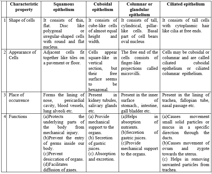
Depending upon the shape and function of the cells, the epithelial tissues are classified as follows :
A. Squamous epithelium : thin flattened cell present in outer lining of skin, blood vessels etc.
B. Cuboidal epithelium : Present in outer lining of kidney tubeles, tube like cells.
C. Columnar epithelium : – long narrow cell. In stomach it form microvilli which increases the absorptive surface of cell.
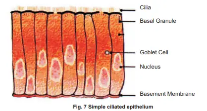
D. Ciliated epithelium :Cells are columnar but war cilia on their surface. Present in respiratory passage, fallopian tube etc
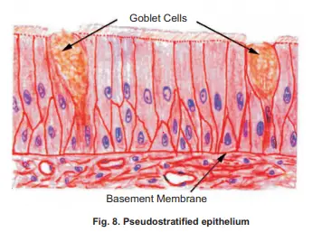
E. Pseudostratified epithelium – Cell, single layered but appear multilayered.
F. stratified epithelium the cells are present in number of layer and form tough and more impervious barrier.
G. Grandular epithelium – An aggregate of grandular cells form a multicellular gland may be present amongst the epithelial cell. For example. exocrine and endocrine gland.
Differences between different types of epithelial tissues : Muscular tissue :
Muscular tissue constitutes all the muscles of the body of an animal.
Muscle cells are elongated and large sized, so they are called muscle fibres.
Muscle cells are typically arranged in parallel arrangement allowing them to work together effectively.
This tissue is responsible for movement in our body. Muscles contain special proteins called contractile proteins, which contract and relax to cause movement.
On the basis of their location, structure and function, there are following three types of muscle fibres :
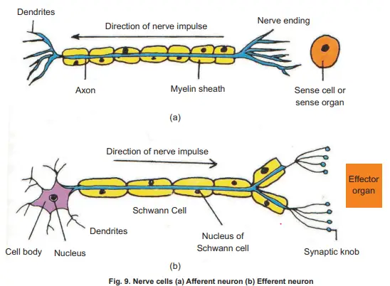
A tissue which is specialized to transmit messages in our body is nervous tissue. Brain, spinal cord and nerves are all composed of nervous tissue.
Nervous tissue contains highly specialized unit cells called nerve cells or neurons.
These cells are specialized for the conduction of impulse over great distance at great speed.
A neuron consists of a cell body (cyton or soma) with a nucleus and cytoplasm, from which long thin hair-like parts arise called dendrons.
Dendrons further branched out to form dendrites. From the distal part of cyton arises a very long process called axon.
Functions :
The nervous tissue is responsible for the reception and transmission of information between different parts of the body.
The dendrites receive impulses and the axon takes impulses away from the cell body.
Connective Tissue
The connective tissue is specialized to connect and anchor various body organs. As such, it connects one bone with another and a bone with a muscle.
Three components are present in all the connective tissues. These are intercellular medium, connective tissue cells and fibres.
The cells of connective are loosely spaced and embedded in an intercellular matrix. The matrix may be jelly like, fluid, dense or rigid.
The nature of matrix decides the function of connective tissue.
General functions :
Connective tissue binds other tissues together in the organs.
Connective tissue also provides the structural framework and mechanical support to different tissues.
It is also concerned with body defence, fat storage, repair etc.
The main functions of connective tissue are binding, supporting and packing together different organs of the body.
Types of connective tissue :
In animals, there are following five types of connective tissues :
1. Areolar (Loose) connective tissue
2. Dense connective tissue
3. Adipose connective tissue
4. Skeletal tissue
5. Fluid connective tissue
Areolar (loose) connective tissue :
It is loose and cellular connective tissue. Its matrix consists of two kinds fibres : white collagen fibres and yellow elastic fibres.
Areolar connective tissue is found between the skin and muscles, around blood vessels and nerves and in the bone marrow.
It fills the spaces between different tissues and organs, hence called packing tissue.
Functions :
It acts as supporting and packing tissue between organs lying in the body cavity.
It provides rapid diffusion of oxygen and nutrients from blood vessels.
It helps in repair of tissues after an injury.
It helps in fighting foreign antigen and toxin.
Dense connective tissue
It is a fibrous connective tissue. It is characterized by ordered and densely packed collection of fibres and cells.
It is the chief component of ligaments and tendons.
Ligaments : These are elastic structures made up of yellow elastic fibrous tissues which connect bone to another. It has considerable strength. Ligaments contain very little matrix. Ligaments strengthen the joint and they permit normal movement but prevent over-flexing or over-extension. Sprain is caused by excessive pulling (stretching) of ligaments.
Tendons : Te dons are cord like, strong inelastic structures that join skeletal muscles to bones.
They are composed of white collagen fibrous tissue.
It has great strength, but its flexibility is limited.
Adipose tissue
It consists of large number of oval and rounded adipose cells (adipocytes) filled with fat globules.
The adipose tissue is abundant below the skin, between the internal organs (e.g., around the kidney) in yellow bone marrow.
Functions :
It serves as a fat reservoir.
Adipose tissue acts as food reservoir by storing fat.
It acts as an insulator and regulates body temperature.
Stem Cells
Most cells after repeated division tend to differentiate into cells with specialized function.
Cells with the ability to differentiate as well as self-renewal are called stem cell.
Depending on the potential to differentiate into different types of cell stem cells are classified as
(a) totipotent-can differentiate into all cell type like zygote.
(b) pluripotent -can differentiate into many cell type but not all. For example-placenta cannot be formed by them.
(c) Multipotent – limited number of cell type for example Bone marrow in adult.
(d) Unipotent – only one cell type.
Depending on the source they are derived from stem cells are of two types. Embryonic stem cell
Derived from inner cell mass of blastocyst when embryo is 4-8 cells old, these are pluripotent but can be made to differentiate in about 200 different cells . For example-mouse embryonic stem cells, muscle cell lung cell, liver cell etc.
Embryonic stem cell have tremendous potential to generate organs and tissues for replacement of damaged or diseased organ or tissues (regenerative medicines).
Through stem cell therapy tissue generated from pluripotent cell may be used to replace damaged muscle of chronic heart patient etc.
In some countries use of ES has been banned as it causes destruction of human embryo.
Adult stem cell
Found in developed organism and can be obtained from fetal card blood and bone marrow these are multipotent and can be changed into pluripotent for developing any type of tissue for medical development.
Biodiversity and its conservation
All forms of life be the bacteria, plants or animals are represented by a number of species. Within each species there are numbers varieties of strain or types. This variety constitute biodiversity.
Importance
It provides human being with food fibres medicine and life sustaining oxygen. It is also important for recycling of waste and regulation of global climate. It makes the ecosystem more productive, stable and silent. Loss of biodiversity
Due to many men made changes such as pollution deforestation, dam construction there is destruction of habitat as a result flora and fauna of that habitat is also getting destroyed leading to loss of biodiversity. Efforts taken to undertake, maintain or present constitute biodiversity conservation.
What are biological hotspots?
A biodiversity hotspot is a biographic region that is both a significant reservoir of biodiversity and is threatened with destruction.
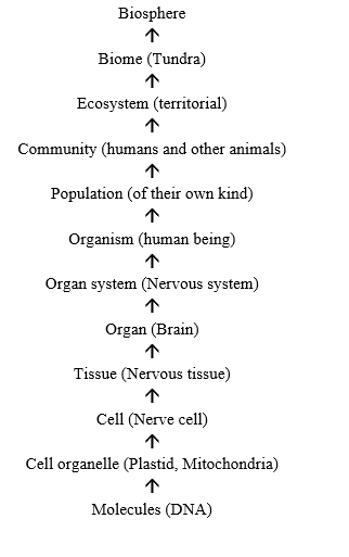
Organization
Biologist organized living creatures and component into a biological hierarchy which is as follow :