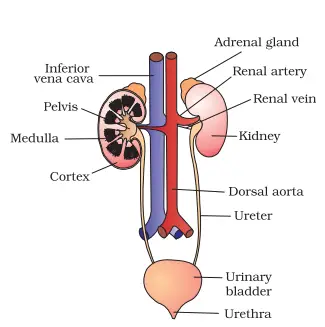
It consists of a pair of Kidneys, a pair of ureters, Urinary bladder and Urethra.
The Kidneys are located in the abdominal cavity, situated below the level of last thoracic and third lumbar vertebra close to the dorsal inner wall of abdominal cavity.
Each kidney is been shaped reddish brown.
The right kidney is lower and smaller than left kidney because the liver takes up much space on the right side.
From each kidney, one ureter arises, and the two Ureters open obliquely into the Urinary bladder, which is a hollow, muscular sac-like structure that stores urine.
Urethra is the membranous tube that arises from the neck of the bladder and conduct the urine to the exterior.
Structure of kidney:
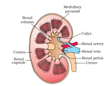
The outer surface of each kidney is convex, and the inner surface is concave, where it has notch called hilum.
Each kidney has three protective covering that is renal fascia (outermost layer), the adipose layer and then renal capsule (innermost layer).
Inside the kidney, outer cortex and inner medulla region are present. The medulla is divided into few conical masses call Medullary Pyramids.
The cortex extend in between the pyramids as renal column is called Column of Bertini.
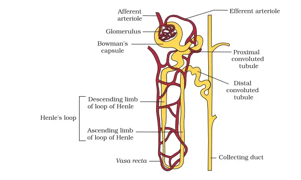
Each kidney has nearly 1 million Complex structures called nephrons which are the functional unit.
Structure of Nephron:
Each nephron consists of two parts that is Glomerulus and Renal tubule.
Glomerulus along with Bowman's capsule is called Malpighian body or Renal corpuscles.
Glomerulus is a tuf of capillaries (bunch) formed by afferent arterioles (branch of renal artery).
Bowman's capsule is a double walled cup like structure that surrounds the glomerulus.
Just below the glomerulus, the tubule has three distinct regions:
Proximal convoluted tubule (PCT)
Loop of Henle's
Distal convoluted tubule (DCT)
Proximal convoluted tubule:
Behind the neck the tubule continue to form a highly coiled network and is restricted to the cortical region of kidney.
Henle's loop:
It is a quite narrow and U-shaped hair pin like loop with a descending limb that ends into the medulla and an ascending limb that coarse back to the cortex.
Distal convoluted tubule:
The ascending limb on entering the cortex become highly coiled distal convoluted tubule. It then continues as short straight collecting tubule that joins the collecting duct. Each collecting duct receives the collecting tubules of number of nephrons.
Types of Nephron:
There are two types of Nephron, based on their position in the Kidney.
Cortical nephron - In majority of nephrons, the loop of henle is too short and extends only very little into the medulla, such nephrons are called cortical nephron.
Juxtamedullary nephron: In some of the nephron, the loop of henle is too long and runs deep into the medulla. These nephrons are called juxtamedullary nephron.
Urine formation:
Urine formation involves three major steps:
Glomerular filtration
Tubular reabsorption/ selective reabsorption
Tubular secretion
Glomerular filtration:
The first step in urine formation is the filtration of blood which is carried out by the glomerulus and is called glomerular filtration. On an average 1100 to 1200 ml of blood is filtered by the kidney per unit that is one fifth of the blood pumped by the ventricles of heart per minute.
The glomerular capillary blood pressure causes filtration of blood through three layer that is endothelium of glomerular blood vessels, epithelium of bowman's capsule and the basement membrane between these two layers.
The epithelial cells of bowman's capsule are called podocytes and arranged in such a manner so as to leave some minute space called filtration slits.
Blood is filtered so finally through these membranes that almost all the constituent of Plasma except proteins pass on the lumen of bowman's capsule. Therefore, this process is considered as of ultrafiltration.
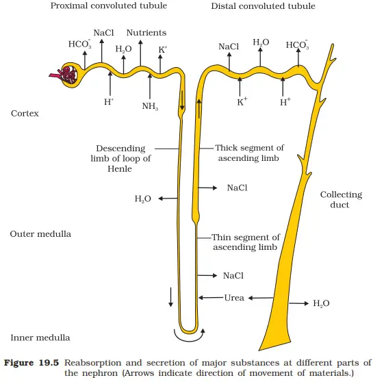
Tubular reabsorption:
A comparison of the volume of filtrate formed by per day that is 180 litre with that of urine released that is 1.5 litre, suggest that nearly 99% of the filtrate has to be reabsorbed by the renal tubule. This process is called selective reabsorption.
Water and urea reabsorbed by passive transport, glucose and amino acids are reabsorbed by active transport. The reabsorption of sodium Ions (Na+ occur both by active and passive transport.
Tubular secretion
Certain chemicals in the blood that are not removed by filtration from the glomerular capillary are removed by the process of tubular secretion. It helps in the maintenance of ionic and acid base balance of the body fluid by removing ions like H+, K+, NH4+ etc.
Glomerular filtration rate (GFR) and Autoregulatory mechanism of GFR:
It refers to the quantity of filtrate formed by the kidney per minute. It is about 125 ml per minute or 180 litre per day in a healthy human individual.
Auto regulatory mechanism of GFR:
The kidneys have an inbuilt mechanism for the regulation of GFR. Juxtaglomerular apparatus (JGA) is a specialised cellular apparatus located where the DCT passes close to the bowman's capsule between the afferent and efferent arterioles.
A fall in GFR activates the cells of JGA to release renin which increases the glomerular blood flow thereby GFR back to normal.
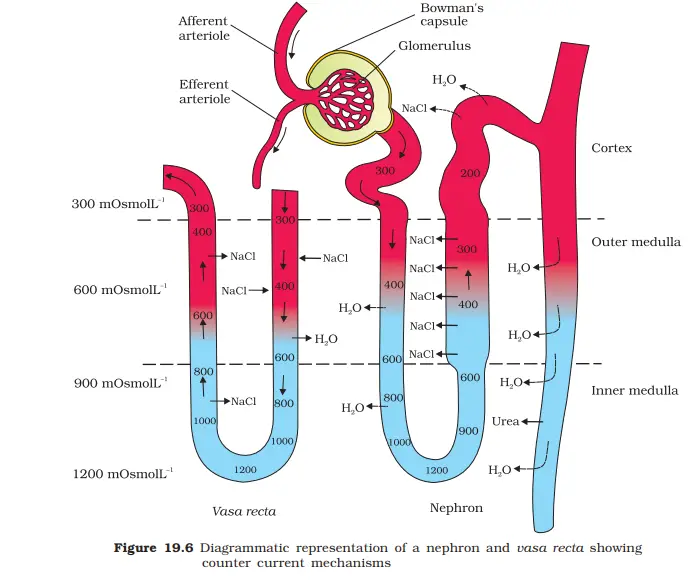
Counter-current mechanism
The loop of Henle and the vasa recta are responsible for concentrating the filtrate (urine). They constitute a mechanism called counter-current mechanism. The flow of filtrate in the two limbs of the loop of Henle is in opposite direction and the flow of blood in the two limbs of Vasa recta is also in the opposite direction and hence, form a counter-current system.
The two factors responsible for increasing the osmolarity towards the medullary interstitium are- (i) The proximity between loop of henle and Vasa recta (ii) The counter-current system in them.
The osmolarity in the cortex is about 300m.osmol/L and that in the medulla about 1200m.osmol/L. This gradient is maintained by NaCl and urea. The interstitial gradient of NaCl is maintained by the loop of Henle. Urea is added to the interstitial fluid of medulla by its diffusion from the collecting duct, if it re-enter the ascending limb by diffusion. The counter-current mechanism thus, help to maintain a concentration gradient between the medullary interstitium and urinary tubule.
As the filtrate moves in the collecting duct pass the interstitial fluid, water moves out of tubule by osmosis and urine become concentrated.
Regulation of kidney function:
By hypothalamus:
When there is a change in osmolarity, and volume of blood and the blood fluid, the osmoreceptors are stimulated which stimulate the hypothalamus to release anti-diuretic hormone (ADH) for vasopressin from the posterior pituitary.
ADH signal the DCT, collecting tubule and collecting duct to reabsorb water from filtrate to prevent diuresis.
It also causes constriction of blood vessels and increases blood pressure which increases GFR.
By Juxtaglomerular Apparatus:
JGA operates through multi hormonal system called Renin Angiotensin- aldosterone system (RAAS). When glomerular blood flow decreases, JGA releases renin in which converts angiotensinogen in the blood into angiotensin I and then angiotensin II.
Angiotensin II is a powerful vasoconstrictor and increases glomerular blood pressure and maintain GFR. it also activate the adrenal cortex to release aldosterone which stimulates reabsorption of Na+ and H2O from DCT which leads to gfr back to normal.
By atrial wall of heart:
An increase in the blood flow and pressure to the atria of the heart causes the release of atrial natriuretic factor (ANF) which causes vasodilation and increases the blood pressure.
ANF functions antagonistically to the renin angiotensin mechanism by inhibiting the release of renin.
Micturition
It is the act of voiding or releasing urine from the urinary bladder. This is accomplished by simultaneous contraction of smooth muscles of urinary bladder wall and relaxation of the skeletal muscle of sphincter of the bladder into the urethra.
Urine:
An adult human excretes about 1- 1.5 litre of urine per day. The urine has straw yellow colour and slightly acidic (pH 6) with the characteristic odour. It contains urea, creatinine and very little amount of ammonia and uric acid.
Glucose (glycosuria) and Ketone bodies (ketonuria) are found in the urine of the patient of diabetes mellitus.
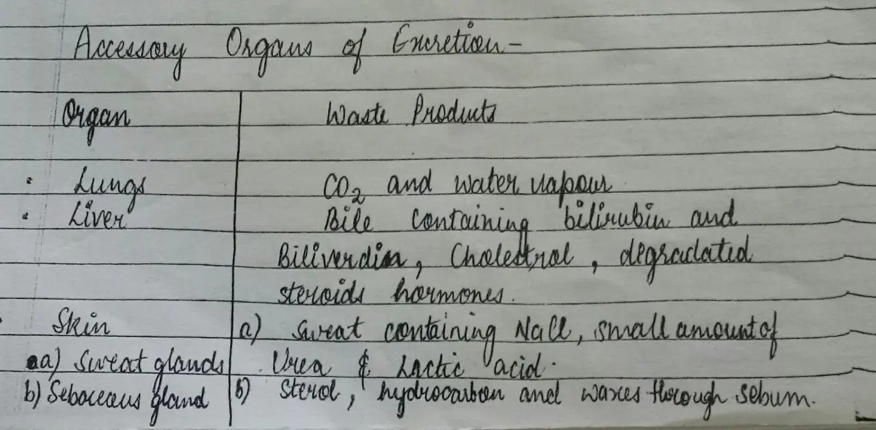
Accessory Organs of Excretion
Disorders of Excretory System:
Malfunctioning of Kidneys can lead to accumulation of Urea in blood, a condition called Uremia which is highly harmful and may lead to kidney failure. In such patients urea can be removed by a process called hemodialysis.
Kidney transplantation is the ultimate method in the correction of acute renal failure that is kidney failures. A functioning kidney is used in transplantation from a Donor, preferably a close relative to minimise its chances of rejection by the immune system of the host.
Renal calculi: Stone or insoluble mass of crystallized salts (oxalate etc) formed within the kidney.
Glomerulonephritis: Inflammation of glomeruli of kidney.