Human-being like most of mammals are unisexual or dioecious (sexes are separate).
Each human-being has only either male or female gonads (primary sex organs) reproductive ducts and accessory genital structures (secondary sex organs).
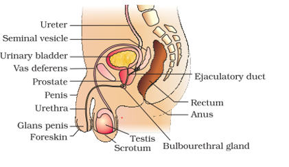
Human Male Reproductive System
It is concerned with sperm formation. It is located in the pelvis region. It consists of
The primary sex organs i.e. pair of testes.
The secondary sex organs i.e. duct system and associated glands.
The external genitalia.
Testes
There are one pair, small- sized, oval- shaped, pinkish in color, testes located outside the abdominal cavity within a pouch called scrotum.
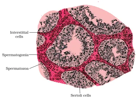
The testes are covered by a dense covering called tunica albuginea.
Internally a testis is divided into about 250 compartments called testicular Lobules.
Each lobule contains 1-3 highly coiled seminiferous tubules in which sperm are produced.
Each seminiferous tubule is lined on its inside by 2 types of cells called male germ cell (spermatogonia) and Sertoli cells.
The male germs cells undergo meiotic division finally leading to sperm formation while Sertoli cells provide nutrition to the germ cell.
The region outside the seminiferous tubules is called interstitial spaces containing small blood vessels and interstitial cells called Leydig cells.
Leydig cells synthesize and secrete male sex hormones called androgens of which testosterone is the principle one.
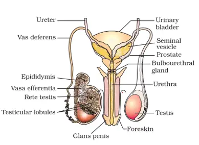
Duct System
The male sex accessory ducts include rete testes, vasa efferentia, epididymis and vasa deferens.
The seminiferous tubule of the testes opens into the vasa efferentia through the rete testes. The vasa efferentia leave the testes and open into epididymis located along the posterior surface of such testes.
The epididymis leads to vasa deferens that ascends to the abdomen and loop over the urinary bladder.
It receives a duct from seminal vesicles and opens into the urethra as the ejaculatory duct.
It passes through prostate grand and joins the urethra.
The urethra originates from the urinary bladder and extends through the penis through its external opening called urethral meatus.
Accessory Glands
The main accessory glands include
A pair of seminal vesicles
A prostate gland
A pair of bulbourethral glands (Cowper’s gland) ( lubrication of penis)
Their secretions are collectively called as seminal plasma which is rich in fructose, calcium and certain enzymes. Exteranl Genitalia
Penis is the external genitalia in human males.
It is made up of special erectile tissue which helps in the erection of penis to facilitate insemination.
The enlarged tip of the penis is called glans penis which is covered by foreskin or prepuce.
Diagrammatic sectional view of male pelvis showing reproductive system
Diagrammatic view of male reproductive system(part of testis is open to show inner details)
Major functions of Male Reproductive system
Spermatogenesis by germ cells of seminiferous tubules.
Secretion of male hormone, testosterone
Transfer of sperm into vagina of females during copulation.
Extra Notes:
Testes
They are involved in spermatogenesis and secretion of testosterone. Epididymis
It is involved in storage, nutrition and physiological maturation of the sperms. It also show peristalsis and segmentic contraction to move the sperm. Vasa efferentia
Conduction of sperms by peristalsis of its highly muscular coat. Urethra
Conduction of sperm, secretion of accessory reproductive glands and urine. Diagrammatic sectional view of seminiferous tubule
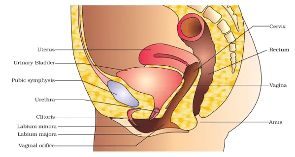
Female Reproductive System
It is concerned with formation of Ova, fertilization, Foetus development and childbirth.
It is located in the pelvis region.
It consists of:
The primary sex organs; pair of ovaries.
The secondary sex organs; i.e. the duct system consisting of pair of fallopian tubes, a uterus, cervix and vagina.
The external genitalia
Mammary glands.
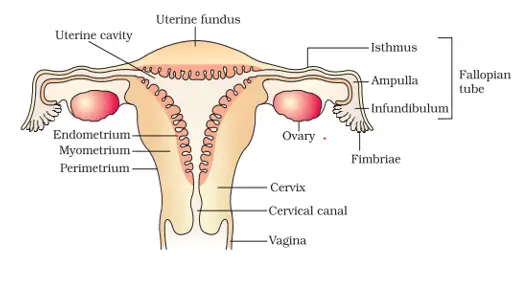
Ovary
These are one pair, small- sized, almond shaped structures present in the pelvis, one on either side of the uterus.
Ovary is covered by a thin epithelium which encloses the ovarian stroma (cavity).
The stroma is divided into two regions:
A peripheral cortex
An inner medulla
Fallopian Tubes
These are one pair, long (10-12) cm, ciliated, muscular and tubular structure which extend from the periphery of ovaries to uterus.
The part of the fallopian tube, closer to the ovary, is funnel-shaped and is called infundibulum.
It’s edges have finger-like projections called Fimbriae which help in collection of ova after ovulation.
The infundibulum leads to the wider part of the fallopian tube called ampulla.
The last part of the fallopian tube that joins the uterus is narrow and is called isthmus.
Uterus
It is a pear-shaped muscular structure attached to the pelvic wall and supported by ligaments.
The wall of the uterus has 3 layers of tissues. The outermost / external layer is the thin membrane's perimetrium, the middle thick layer of smooth muscles is called myometrium and the innermost glandular layer is endometrium which lines the uterine cavity and undergoes cyclic changes during menstrual cycle.
The uterus opens into the vagina through a narrow cervix. The cavity of the cervix is called the cervical canal.
Vagina
It is a muscular tube-like structure that opens to the outside.
The vagina is partially covered by a membrane called himen.
Diagrammatic sectional view of female pelvis showing reproductive system
Diagrammatic sectional view of the female reproductive system
External Genitalia
The female external genitalia include mons pubis, labia majora, labia minora, hymen and clitoris.
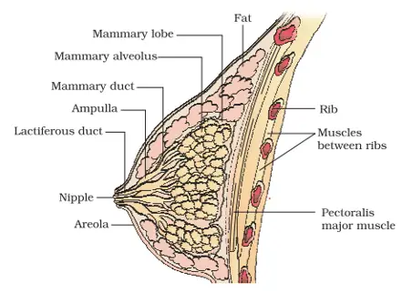
Mammary Glands (Breast)
A human female has a pair of functional mammary glands that consists of glandular tissue and variable quantity of fat.
The glandular tissue is divided into 15-20 mammary lobes containing clusters of cells called alveoli which open into mammary tubules. (secrete milk which is stored in cavities (lumen) of alveoli)
The mammary tubules of each lobe join to a mammary duct.
Several mammary ducts join to form a wider mammary ampula which is connected to the lactiferous duct through which milk comes out. (sucked out)
A diagrammatic sectional view of Mammary gland
Major Functions
oogenesis by the germ cells of the ovary.
fertilization of gametes to form zygote.
Implantation followed by prenatal growth of embryo.
Parturition
Note: Ovary:
They are concerned with formation of ova and secretion of female sex hormones; i.e. estrogen and progesterone. Fallopian Tubes: (ovary duct)
It is involved in the conduction of ovum or zygote towards the uterus by peristalsis and ciliary action. It is also a site of fertilization. Uterus (womb):
It is the site of implantation and foetal growth during pregnancy. It also takes part in the placenta formation. Vagina:
Serves as the birth canal during parturition. It also acts as a copulation canal as it receives the sperm during copulation.
Gametogenesis
It refers to the process of formation of gametes for sexual reproduction.
The primary sex organs- the testes in males and ovalies in female produce gametes i.e. sperm and ovum respectively by the process called gametogenesis.
Gametogenesis is of two types
Spermatogenesis
Oogenesis
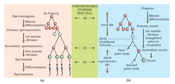
Spermatogenesis
It is the process of formation of spermatozoa (sperms) in the testes, the process starts at puberty.
The spermatogonial cells present on the inner wall of seminiferous tubules multiply by mitotic division.
Some of the spermatocytes called primary spermatocytes periodically undergo meiosis.
A Primary spermatocyte completes the first meiotic division leading to formation of two equal, haploid cells secondary spermatocytes.
The secondary spermatocytes undergo second meiotic division to produce 4 equal haploid spermatids.
The spermatids are transformed into spermatozoa (sperm) by the process called spermiogenesis.
After spermatogenesis sperm heads become embedded in the sertoli cells and are finally released from the seminiferous tubule by the process called spermiation.
Hormonal Control of Spermatogenesis
In human males, spermatogenesis starts only at the age of puberty due to increased secretion of Gonadotropin Releasing Hormone (GnRH) by hypothalamus of the brain.
The increased level of GnRH act on the anterior pituitary and stimulates the selection of two gonadotropins i.e. Luteinizing hormone (LH) and follicle stimulating hormone (FSH).
It acts on the leydig cells and stimulates them to secrete testosterone: stimulate the process of spermatogenesis.
FSH acts on sertoli cells and stimulates secretion of some factors which help in spermeiogenesis.
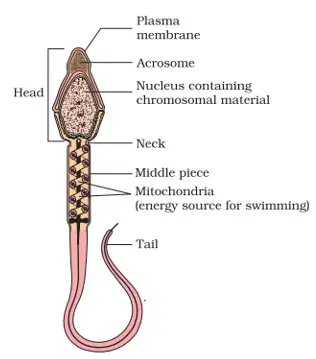
Structure of Spermatozoa (sperm):
The whole body of spermatozoa is enveloped by plasma membrane.
A human sperm is composed of 4 parts:
Head, Neck, middle piece, and tail. Head:
The sperm head contains an elongated haploid nucleus, the interior portion of which is covered by a cap-like structure acrosome. Neck:
Neck contains two centrioles; proximal and a distal centriole. Middle piece:
Middle piece contain a number of mitochondria that provide the energy for the motility of sperm. Tail:
It consists of axial filaments surrounded by plasma membrane. Structure of Sperm
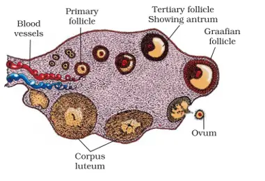
Oogenesis:
It is the process of formation of female gametes or ova in the ovary.
Oogenesis is initiated during embryonic development in the female foetus.
The oogonia (about a million) are formed in the ovary of the foetus of 25 weeks; no more oogonia are formed after it.
The oogonial cells start division and enter into prophase I of the meiosis I division and get temporarily arrested at that stage called primary oocyte.
The primary oocyte becomes surrounded by a layer of granulosa cells and is called the primary follicle.
The primary follicle becomes surrounded by more granulosa cells and a thecal layer at this stage they are called secondary follicle.
Soon secondary follicle develops a fluid filled antral cavity called antrum and now it is called tertiary follicle.
As the tertiary follicle continued its development the primary oocyte completed its meiosis first and formed a large cell; the secondary oocyte and a tiny cell, the first polar body.
The secondary oocyte becomes changed into a mature follicle called graafian follicle.
The secondary oocyte starts its II meiotic division, but it is suspended in metaphase II until a sperm enters.
The secondary oocyte forms a new membrane called zona pellucida surrounding it.
The graafian follicle now ruptures to release the secondary oocyte from the ovary by the process called ovulation.
Diagrammatic Section view of ovary
Schematic representation of (a) Spermatogenesis; (b) Oogenesis
Significance of Spermatogenesis
Produce haploid sperms.
Crossing over may occur during meiosis 1 so producing variation.
Proves evolutionary relationship.
Significance of oogenesis
Produce haploid ovum by releasing 2 or3 haploid polar bodies.
Most of cytoplasm retains in the functional ovum.
Variation may appear due to crossing over during meiosis I.
Process evolutionary reaction.
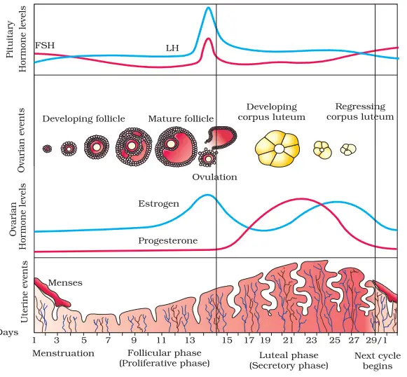
Menstrual Cycle
The reproductive cycle in the female primates eg. Monkey, apes and human-being is called menstrual cycle.
The cyclic changes that occur in the reproductive organs of female primates continue the menstrual cycle.
In human females, menstruation is repeated at an average interval of about 28/29 days and the cycle of events starting from one menstruation till the next one is called menstrual cycle.
The first menstruation begins at puberty and is called menarche.
One ovum may released during the middle of each menstrual cycle.
The Events in a Menstrual Cycle can be studied under 4-phases: Menstrual Phases:
The cycle starts with this phase at the menstrual flow (menstruation) lasts for 3-5 days.
It results due to the breakdown of endometrial lining of the uterus and its blood vessels along with the unfertilized ovum.
Follicular Phase/Proliferative Phase
In this phase, the primary follicle in the ovary grows and becomes a fully mature graafian follicle.
The endometrium of the uterus is regenerated by proliferation of its cell.
These changes are due to an increased level of pituitary (FSH and LH) and ovary hormones (estrogen).
FSH controls the follicular phase; it stimulates the growth of follicle and secretion of estrogen by the growing follicle.
Both FSH and LH reach their peak level in the middle of the cycle (about 14th day).
Ovulatory phase:
The peak level of LH (called LH surge) induces the rupture of the mature graafian follicle and thereby releases the ovum, this process is called ovulation. Luteal Phase/ Secretory Phase
During this phase the ruptured follicle is transformed into corpus luteum.
It secretes large quantities of progesterone.
The endometrium thickens further and their gland secrete a fluid into the uterus.
In the absence of fertilization, corpus luteum degenerates and this causes disintegration of the endometrium leading to menstarvation.
Diagrammatic presentation of various events during a menstrual cycle
Menopause:
The menstrual cycle ceases at the age of about 45-50 years, it is called menopause.
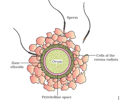
Fertilization
Fertilization also called syngamy involves the fusion of haploid male and female gametes to form diploid zygote.
Fertilization in human beings is internal.
Fertilization takes place in the place of the fallopian tube of the female.
During copulation, the semen is transferred into vagina (insemination).
The motive sperm moves through the cervix, enters the uterus and reaches the ampulla isthmic junction(fallopian tube) where fertilization takes place.
A sperm comes in contact with the zona pellucida layer of ovum and induces changes in the membrane to block the entry of other sperms-(dissolves the membrane).
The secretion of the acrosome helps the sperm enter into the cytoplasm of its own through the zona pellucida and the plasma membrane.
The entry of sperm induces the completion of II meiotic division of the secondary oocyte,that results in the formation of a haploid ootid, hence a second polar body.
Soon ,the haploid nucleus of sperm and that of the ovum fuse together to form a diploid zygote.
Ovum surrounded by few sperms
Implantation
The process of attachment of blastocyst (mammalian blastula) on the endometrium of the uterus is called implantation. Cleavage
The zygote undergoes successive mitotic division called cleavage,as it moves through the isthmus of the fallopian tube towards the uterus.
The daughter cells are called blastomere. Morula:
At the 16th celled stage, the embryo is a solid sphere and is called morula.
Blastocyst
The morula continues to divide and transform into blastocyst.
The blastomeres in the blastocyst are arranged in an outer layer called profoblast, an inner group of cells attached to profoblast called the inner cell mass.
Transport of ovum, fertilisation and passage of growing embryo through fallopian tube
Implantation:
The trophoblast layer gets attached to the endometrium.
The cells of the endometrium divide rapidly and covers the blastocyst.
So the blastocyst gets embedded in the endometrium. This process is called implantation.
The cells of inner cell mass differentiate to form the embryo proper.
Gastrulation is a process by which blastocyst change into gastrula with 3 primary germ layers.
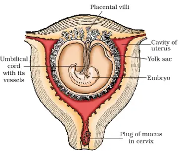
Pregnancy and Embryonic Development
After implantation finger-like projections appear on the trophoblast called chorionic villi which are surrounded by the uterine tissue and maternal blood.
The chorionic villi and the uterine tissue become interdigitated to form the structural and functional unit called placenta.
The placenta facilitates the supply of O2 and nutrients to the embryo, also removal of CO2 and excretory/ waste material produced by the embryo.
The placenta secretes hormones like Human chorionic gonadotrophins (hCG), estrogen and progesterone, that are necessary to maintain pregnancy.
Umbilical cord, the structure that connects the placenta with the foetus is formed.
Simultaneously the inner mass differentiate into an outer layer called ectoderm and an inner layer called endoderm.
A middle layer called mesoderm appears b/w ectoderm and endoderm.
These primary germ layers give rise to all the tissues and organs to the adult.
The human foetus within the uterus NOTE:
Organogenesis includes the formation of a specific organ system of the body from 3 primary germ layers of Gastrula.
Parturition
It is an expelling of the fully formed young one from the mother’s uterus after the gestation period i.e. of about 9.5 months.
Parturition is induced by a complex neuroendocrine mechanism.
The signals for parturition originate from the fully developed foetus and the placenta which induce mild uterine contraction called foetal ejection reflex.
This triggers the release of oxytocin from the maternal pituitary.
Oxytocin induces stronger contraction of the uterine muscles which ultimately leads to the expulsion of the baby from the uterus through the birth canal.
It is followed by the expulsion of the placenta and the remains of the umbilical cord.
Lactation
Lactation involves the synthesis, secretion and ejection of milk.
The mammary glands of the female undergo differentiation during pregnancy ( under the influence of hormones like prolactin and progesterone) and produce milk towards the end of pregnancy by the process called lactation.
The milk that comes out from the mammary glands of the mother just after childbirth is called colostrum. It is rich in nutrients and certain antibody (IgA) for the baby.
SIGNIFICANCE OF FERTILIZATION
It restores the diploidy (2n=46 in human beings) in the zygote. Fertilization membrane prevent polyspermy.
It combines the characters of two parents and introduces variation so helps in evolution.
Sex- chromosome of sperm is either X or Y and help in sex-determination.